. 2010 Jan;6(2):256-61.
doi: 10.1002/smll.200901672.

Ultrasmall near-infrared non-cadmium quantum dots for in vivo tumor imaging

Affiliations

Ultrasmall near-infrared non-cadmium quantum dots for in vivo tumor imaging

Jinhao Gao et al. Small. 2010 Jan.

Abstract

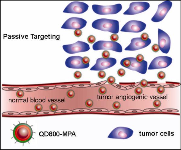
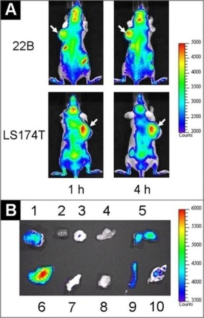
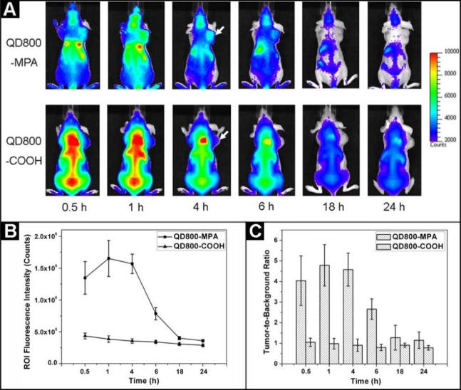
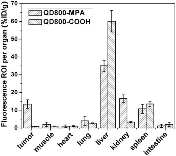
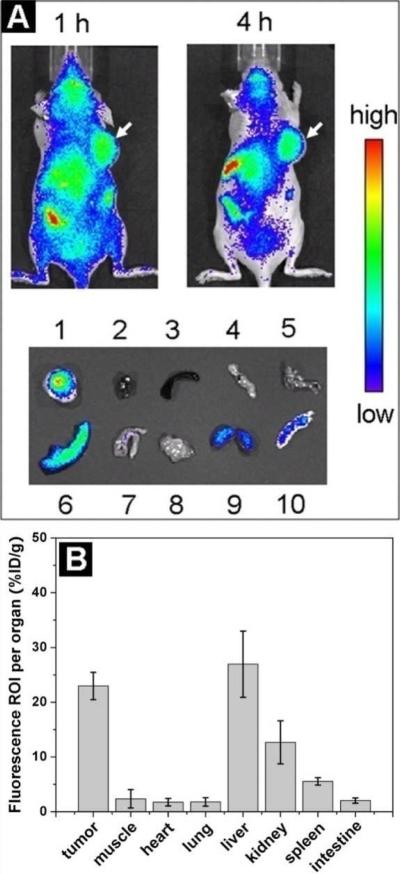
The high tumor uptake of ultrasmall near-infrared quantum dots (QDs) attributed to the enhanced permeability and retention effect is reported. InAs/InP/ZnSe QDs coated by mercaptopropionic acid (MPA) exhibit an emission wavelength of about 800 nm (QD800-MPA) with very small hydrodynamic diameter (<10 nm). Using 22B and LS174T tumor xenograft models, in vivo and ex vivo imaging studies show that QD800-MPA is highly accumulated in the tumor area, which is very promising for tumor detection in living mice. The ex vivo elemental analysis (Indium) using inductively coupled plasma (ICP) spectrometry confirm the tumor uptake of QDs. The ICP data are consistent with the in vivo and ex vivo fluorescence imaging. Human serum albumin (HSA)-coated QD800-MPA nanoparticles (QD800-MPA-HSA) show reduced localization in mononuclear phagocytic system-related organs over QD800-MPA plausibly due to the low uptake of QD800-MPA-HSA in macrophage cells. QD800-MPA-HSA may have great potential for in vivo fluorescence imaging.


Figures

Scheme 1
Scheme 1
The structure of QD800-MPA and the illustration of the passive tumor targeting of QD800-MPA in tumor model.
Figure 1
Figure 1
In vivo and ex vivo fluorescence imaging by the tail vein injection of QD800-MPA. (A) In vivo NIR fluorescence imaging of 22B tumor-bearing mice and LS174T tumor-bearing mice (arrows) at 1 h and 4 h. (B) Representative ex vivo imaging at 4 h time point. 1, 22B tumor; 2, heart; 3, pancreas; 4, intestine; 5, kidney; 6, liver; 7, skin; 8, muscle; 9, spleen; 10, lung.
Figure 2
Figure 2
(A) In vivo NIR fluorescence imaging of 22B tumor-bearing mice (arrows) injected with QD800-MPA and QD800-COOH, respectively. (B) Tumor fluorescence intensity-time and (C) tumor-to-background ratios of mice injected with QD800-MPA and QD800-COOH, respectively. The data were represented as mean ± standard deviation (SD), n = 3/group.
Figure 3
Figure 3
ROI analysis of major organs in ex vivo fluorescence imaging after 4 h p.i. of QD800-MPA and QD800-COOH, respectively.
Figure 4
Figure 4
Overlay of QD800-MPA fluorescence image and FITC-lectin staining image of frozen 22B tumor tissue slices (5 μm in thickness). Arrows point to QD800-MPA particles stayed within tumor blood vessel, while arrowheads indicate extravsated QD800-MPA nanoparticles. Magnification ×200.
Figure 5
Figure 5
(A) In vivo imaging and ex vivo imaging (4 h p.i.) of LS174T tumor-bearing mice (arrows) after the tail vein injection of QD800-MPA-HSA nanoparticles. 1, LS174T tumor; 2, heart; 3, spleen; 4, pancreas; 5, lung; 6, liver; 7, intestine; 8, muscle; 9, kidney; 10, skin. (B) ROI analysis of major organs after 4 h p.i. of QD800-MPA-HSA.

References

    1. Roco MC. Curr. Opin. Biotechnol. 2003;14:337. - PubMed
    1. Whitesides GM. Nat. Biotechnol. 2003;21:1161. - PubMed
    1. Katz E, Willner I. Angew. Chem., Int. Edit. 2004;43:6042. - PubMed
    1. Cai WB, Chen XY. Small. 2007;3:1840. - PubMed
    1. Michalet X, Pinaud FF, Bentolila LA, Tsay JM, Doose S, Li JJ, Sundaresan G, Wu AM, Gambhir SS, Weiss S. Science. 2005;307:538. - PMC - PubMed

Publication types