. 2011 Feb;15(2):296-306.
doi: 10.1111/j.1582-4934.2009.00969.x.

Human ribosomal protein S13 promotes gastric cancer growth through down-regulating p27(Kip1)

Affiliations

Human ribosomal protein S13 promotes gastric cancer growth through down-regulating p27(Kip1)

Xueyan Guo et al. J Cell Mol Med. 2011 Feb.

Abstract

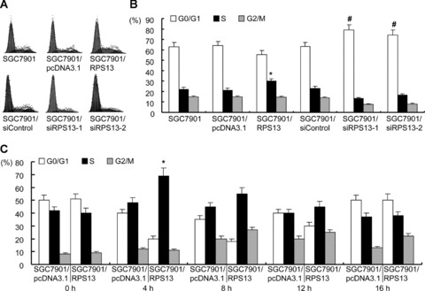
Our previous works revealed that human ribosomal protein S13 (RPS13) was up-regulated in multidrug-resistant gastric cancer cells and overexpression of RPS13 could protect gastric cancer cells from drug-induced apoptosis. The present study was designed to explore the role of RPS13 in tumorigenesis and development of gastric cancer. The expression of RPS13 in gastric cancer tissues and normal gastric mucosa was evaluated by immunohistochemical staining and Western blot analysis. It was found RPS13 was expressed at a higher level in gastric cancer tissues than that in normal gastric mucosa. RPS13 was then genetically overexpressed in gastric cancer cells or knocked down by RNA interference. It was demonstrated that up-regulation of RPS13 accelerated the growth, enhanced in vitro colony forming and soft agar cologenic ability and promoted in vivo tumour formation potential of gastric cancer cells. Meanwhile, down-regulation of RPS13 in gastric cancer cells resulted in complete opposite effects. Moreover, overexpression of RPS13 could promote G1 to S phase transition whereas knocking down of RPS13 led to G1 arrest of gastric cancer cells. It was further demonstrated that RPS13 down-regulated p27(kip1) expression and CDK2 kinase activity but did not change the expression of cyclin D, cyclin E, CDK2, CDK4 and p16(INK4A). Taken together, these data indicate that RPS13 could promote the growth and cell cycle progression of gastric cancer cells at least through inhibiting p27(kip1) expression.


Figures

Fig 1
Fig 1
RPS13 expression in gastric cancer tissues. (A) Western blot analysis of RPS13 protein in gastric tissues. Proteins extracted from gastric cancer (T) and adjacent normal (N) tissues taken from 13 patients were separated by 12% SDS-PAGE and subjected to Western blot analysis using anti-RPS13 and anti-β-actin specific antibodies. Four representative pairs of tissues are presented. (B) Immunohistochemical staining of RPS13 in 24 non-tumour gastric mucosa tissues and 61 gastric cancer tissues. Expression of RPS13 was determined by immunohisto-chemical staining with anti-RPS13 antibody. Shown are representative staining of RPS13 in non-tumour gastric mucosa tissues (a, fundic actively growing gastric epithelia cells display positive staining), well-differentiated gastric carcinoma (b), moderately differentiated gastric carcinoma (c) and poorly differentiated gastric carcinoma (d). Original magnifications: 100×. (C) Percentage of cases showing RPS13 immunoreactivity in 24 normal gastric mucosa tissues and 61 gastric cancer tissues. The immunostaining pattern was scored according to a four-tiered system based on the percentage of immunoreactive tumour cells. *P < 0.01 versus normal tissues.
Fig 2
Fig 2
RPS13 expression in gastric cancer cell lines. RPS13 expression in human gastric cancer cell lines was determined by Western blot analysis with anti-RPS13 antibody. β-actin was probed as loading control. (A) Detection of RPS13 in human gastric cancer cell lines MKN28, MKN45, SGC7901, AGS and immortalized gastric mucosa epithelial cell line GES. (B) Expression of RPS13 in SGC7901 and its derivates. SGC7901 cells were transfected with RPS13 construct (in pcDNA3.1), RPS13-specific siRNA constructs (designated as siRPS13, in pSilencer) or the corresponding control vectors. The transfectants were selected with G418 for 2 months. The mixed cell pools of G418-resistant clones were used for analysing the expression of RPS13 by Western blot.
Fig 3
Fig 3
The growth curves of SGC7901 cell derivates. Cell growth was determined by MTT assays as described in ‘Materials and methods’. Values represent the mean ± S.D. of triplicates in one experiment. Shown are representative of at least three separate experiments. *P < 0.05 versus empty vector controls and SGC7901 cells. #P < 0.01 versus scramble control and SGC7901 cells.
Fig 4
Fig 4
Effects of RPS13 on in vitro tumorigenesis of SGC7901 cells. (A) in vitro colony formation assay. Cells were placed into 60 mm dish and cultured for approximately 2 weeks. The cell colonies were fixed with methanol and visualized with Giemsa staining. The ratio of colony formation was calculated as: (colonies formed/cells seeded) × 100%. (B) Soft agar clono-genic assay. Cells were plated in 0.3% agarose with a 0.5% agarose underlay. Two weeks later, the formed colonies were counted. Values represent the mean ± S.D. of triplicates in one experiment. Shown are representative of at least three separate experiments. *P < 0.01 versus SGC7901 and SGC7901/pcDNA3.1 cells. #P < 0.01 versus SGC7901 and SGC7901/siControl cells.
Fig 5
Fig 5
Effects of RPS13 on in vivo tumori-genesis of SGC7901 cells. Nude mice were injected subcutaneously with 3 × 10⁶ cells into the right upper back. Four weeks later, mice were killed and the transplanted tumours were excised (A) and tumour weight was evaluated (B). *P < 0.05 versus SGC7901/pcDNA3.1 cells. #P < 0.01 versus SGC7901/si Control cells.
Fig 6
Fig 6
Cell cycle analysis of SGC7901 variants. (A) SGC7901 and its derivates in log phase were harvested and subjected to cell cycle analysis. The cell cycle phase distribution was evaluated by FACScan. Shown are representative of four separate experiments. (B) The cell cycle distribution was calculated and expressed as mean ± S.D. of four separate experiments described in (A). *P < 0.01 versus SGC7901 and SGC7901/pcDNA3.1 cells. #P < 0.01 versus SGC7901 and SGC7901/siControl cells. (C) SGC7901/pcDNA3.1 and SGC7901/RPS13 cells were synchronized to the G1/S boundary by double thymidine block and then harvested at 4 hr intervals for 16 hrs after releasing. The cell cycle was evaluated by FACScan and expressed as mean ± S.D. of four separate experiments. *P < 0.01 versus SGC7901/pcDNA3.1 cells.
Fig 7
Fig 7
Effects of RPS13 on p27Kip1 expression in SGC7901 cells. (A) Detection of cell cycle associated proteins by Western blot. Proteins extracted from SGC7901 and its variants were resolved over 12% SDS-PAGE, transferred to nitrocellulose membrane and sequentially blotted with antibodies specific to p27Kip1, cyclin E, CDK2, p16INK4A, cyclin D1, CDK4 and β-actin. (B) in vitro CDK2 kinase assay. CDK2 was immunopre-cipitated from total cell lysates of SGC7901 and its derivates. Immunoprecipitates were employed for in vitro kinase assay using histone H1 as substrate, then resolved over SDS-PAGE and transferred to nitro-cellulose membrane. Phospho-labelled histone H1 was visualized by radioautography (upper panel). Afterward, the membrane was probed for CDK2 to confirm equal loading of kinase assay (lower panel). (C) To assess if RPS13 influenced degradation of p27Kip1, the proteasome inhibitor MG132 was added to the cells 2 hrs before harvest. p27Kip1 was detected by Western blot. (D) Detection of p27Kip1 by real-time RT-PCR. Total RNA extracted from SGC7901 and its variants was subjected to real-time PCR to determine p27Kip1 mRNA level. β-actin was used as normalizer. The histograms indicate the expression of p27Kip1 mRNA relative to the level of β-actin mRNA. Data shown represent the mean ± S.D. of three independent experiments. *P < 0.01 versus SGC7901 and SGC7901/pcDNA3.1 cells. #P < 0.01 versus SGC7901 and SGC7901/siControl cells.

Similar articles

Cited by

References

    1. Denis MG, Chadeneau C, Lecabellec MT, et al. Over-expression of the S13 riboso-mal protein in actively growing cells. Int J Cancer. 1993;55:275–80. - PubMed
    1. Zhang L, Cilley RE, Chinoy MR. Suppression subtractive hybridization to identify gene expressions in variant and classic small cell lung cancer cell lines. J Surg Res. 2000;93:108–19. - PubMed
    1. Kondoh N, Wakatsuki T, Ryo A, et al. Identification and characterization of genes associated with human hepatocellular car-cinogenesis. Cancer Res. 1999;59:4990–6. - PubMed
    1. Ganger DR, Hamilton PD, Klos DJ, et al. Differential expression of metallopanstim-ulin/S27 ribosomal protein in hepatic regeneration and neoplasia. Cancer Detect Prev. 2001;25:231–6. - PubMed
    1. Vaarala MH, Porvari KS, Kyllönen AP, et al. Several genes encoding ribosomal proteins are over-expressed in prostate-cancer cell lines: confirmation of L7a and L37 over-expression in prostate-cancer tissue samples. Int J Cancer. 1998;78:27–32. - PubMed

Publication types

MeSH terms