

Structural bases for stability-function tradeoffs in antibiotic resistance
- PMID: 19913034
- PMCID: PMC2815101
- DOI: 10.1016/j.jmb.2009.11.005
Structural bases for stability-function tradeoffs in antibiotic resistance
Abstract
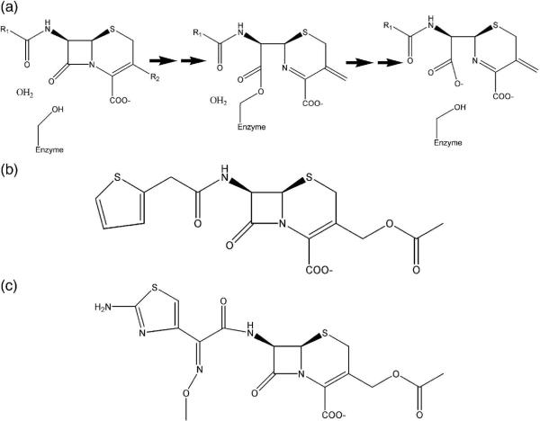
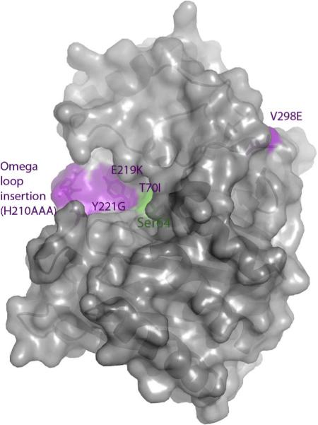
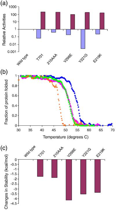
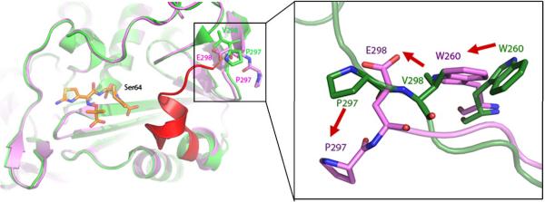
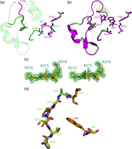
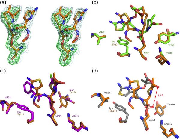
Preorganization of enzyme active sites for substrate recognition typically comes at a cost to the stability of the folded form of the protein; consequently, enzymes can be dramatically stabilized by substitutions that attenuate the size and preorganization "strain" of the active site. How this stability-activity tradeoff constrains enzyme evolution has remained less certain, and it is unclear whether one should expect major stability insults as enzymes mutate towards new activities or how these new activities manifest structurally. These questions are both germane and easy to study in beta-lactamases, which are evolving on the timescale of years to confer resistance to an ever-broader spectrum of beta-lactam antibiotics. To explore whether stability is a substantial constraint on this antibiotic resistance evolution, we investigated extended-spectrum mutants of class C beta-lactamases, which had evolved new activity versus third-generation cephalosporins. Five mutant enzymes had between 100-fold and 200-fold increased activity against the antibiotic cefotaxime in enzyme assays, and the mutant enzymes all lost thermodynamic stability (from 1.7 kcal mol(-)(1) to 4.1 kcal mol(-)(1)), consistent with the stability-function hypothesis. Intriguingly, several of the substitutions were 10-20 A from the catalytic serine; the question of how they conferred extended-spectrum activity arose. Eight structures, including complexes with inhibitors and extended-spectrum antibiotics, were determined by X-ray crystallography. Distinct mechanisms of action, including changes in the flexibility and ground-state structures of the enzyme, are revealed for each mutant. These results explain the structural bases for the antibiotic resistance conferred by these substitutions and their corresponding decrease in protein stability, which will constrain the evolution of new antibiotic resistance.
Copyright 2009 Elsevier Ltd. All rights reserved.
Figures

Similar articles
-
Atomic resolution structures of CTX-M beta-lactamases: extended spectrum activities from increased mobility and decreased stability.J Mol Biol. 2005 Apr 29;348(2):349-62. doi: 10.1016/j.jmb.2005.02.010. J Mol Biol. 2005. PMID: 15811373
-
The Drug-Resistant Variant P167S Expands the Substrate Profile of CTX-M β-Lactamases for Oxyimino-Cephalosporin Antibiotics by Enlarging the Active Site upon Acylation.Biochemistry. 2017 Jul 11;56(27):3443-3453. doi: 10.1021/acs.biochem.7b00176. Epub 2017 Jun 27. Biochemistry. 2017. PMID: 28613873 Free PMC article.
-
Structural bases of stability-function tradeoffs in enzymes.J Mol Biol. 2002 Aug 9;321(2):285-96. doi: 10.1016/s0022-2836(02)00599-5. J Mol Biol. 2002. PMID: 12144785
-
Class D β-lactamases: a reappraisal after five decades.Acc Chem Res. 2013 Nov 19;46(11):2407-15. doi: 10.1021/ar300327a. Epub 2013 Jul 31. Acc Chem Res. 2013. PMID: 23902256 Free PMC article. Review.
-
Structural and Mechanistic Basis for Extended-Spectrum Drug-Resistance Mutations in Altering the Specificity of TEM, CTX-M, and KPC β-lactamases.Front Mol Biosci. 2018 Feb 23;5:16. doi: 10.3389/fmolb.2018.00016. eCollection 2018. Front Mol Biosci. 2018. PMID: 29527530 Free PMC article. Review.
Cited by
-
Cryptic β-Lactamase Evolution Is Driven by Low β-Lactam Concentrations.mSphere. 2021 Apr 28;6(2):e00108-21. doi: 10.1128/mSphere.00108-21. mSphere. 2021. PMID: 33910990 Free PMC article.
-
Deciphering the Evolution of Cephalosporin Resistance to Ceftolozane-Tazobactam in Pseudomonas aeruginosa.mBio. 2018 Dec 11;9(6):e02085-18. doi: 10.1128/mBio.02085-18. mBio. 2018. PMID: 30538183 Free PMC article.
-
Stability-activity tradeoffs constrain the adaptive evolution of RubisCO.Proc Natl Acad Sci U S A. 2014 Feb 11;111(6):2223-8. doi: 10.1073/pnas.1310811111. Epub 2014 Jan 27. Proc Natl Acad Sci U S A. 2014. PMID: 24469821 Free PMC article.
-
Simultaneous enhancement of multiple functional properties using evolution-informed protein design.bioRxiv [Preprint]. 2023 May 9:2023.05.09.539914. doi: 10.1101/2023.05.09.539914. bioRxiv. 2023. Update in: Nat Commun. 2024 Jun 20;15(1):5141. doi: 10.1038/s41467-024-49119-x. PMID: 37214973 Free PMC article. Updated. Preprint.
-
Crystal Structure of OXA-58 with the Substrate-Binding Cleft in a Closed State: Insights into the Mobility and Stability of the OXA-58 Structure.PLoS One. 2015 Dec 23;10(12):e0145869. doi: 10.1371/journal.pone.0145869. eCollection 2015. PLoS One. 2015. PMID: 26701320 Free PMC article.
References
-
- Warshel A, Sussman F, Hwang J-K. Evaluation of Catalytic Free Energies in Genetically Modified Proteins. J. Mol. Biol. 1988;201:139–159. - PubMed
-
- Herzberg O, Moult J. Analysis of the Steric Strain in the Polypeptide Backbone of Protein Molecules. Proteins. 1991;11:223–229. - PubMed
-
- Clackson T, Wells JA. A hot spot of binding energy in a hormone-receptor interface. Science. 1995;267:383–386. - PubMed
-
- Richards FM. The interpretation of protein structures: total volume, group volume distributions and packing density. J. Mol. Biol. 1974;82:1–14. - PubMed
Publication types
MeSH terms
Substances
Associated data
- Actions
- Actions
- Actions
- Actions
- Actions
- Actions
- Actions
Grants and funding
LinkOut - more resources
Full Text Sources
Other Literature Sources
Medical
