. 2009 Nov 11;17(11):1453-64.
doi: 10.1016/j.str.2009.09.010.

Regulation of the protein-conducting channel by a bound ribosome

Affiliations

Regulation of the protein-conducting channel by a bound ribosome

James Gumbart et al. Structure. .

Abstract

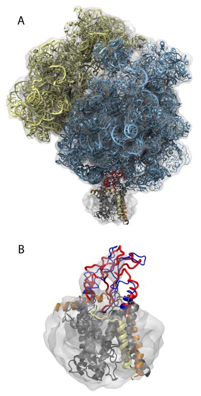
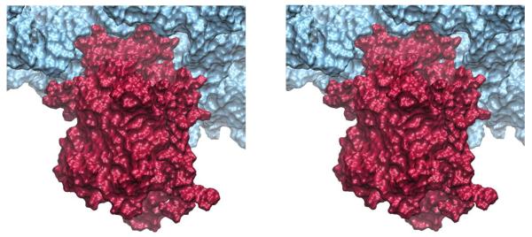
During protein synthesis, it is often necessary for the ribosome to form a complex with a membrane-bound channel, the SecY/Sec61 complex, in order to translocate nascent proteins across a cellular membrane. Structural data on the ribosome-channel complex are currently limited to low-resolution cryo-electron microscopy maps, including one showing a bacterial ribosome bound to a monomeric SecY complex. Using that map along with available atomic-level models of the ribosome and SecY, we have determined, through molecular dynamics flexible fitting (MDFF), an atomic-resolution model of the ribosome-channel complex. We characterized computationally the sites of ribosome-SecY interaction within the complex and determined the effect of ribosome binding on the SecY channel. We also constructed a model of a ribosome in complex with a SecY dimer by adding a second copy of SecY to the MDFF-derived model. The study involved 2.7-million-atom simulations over altogether nearly 50 ns.


Figures

Figure 1
Figure 1
SecYEβ monomer, alone and in complex with the ribosome. (A) SecYEβ viewed from the cytoplasmic side. SecY is in grey with loop 6/7 highlighted in purple, loop 8/9 in red, the plug in blue, and the lateral gate in green. SecE is shown in orange and Secβ in yellow. (B) SecYEβ viewed from the plane of the membrane in the same representation as in (A). (C) Simulation system assumed for the SecYEβ monomer in complex with a ribosome. SecYEβ is shown as in parts A and B. The large subunit of the ribosome is shown in cyan and the small subunit in yellow. The membrane is in yellow with its phosphorus atoms in orange. The surrounding water is indicated as a light blue background. Ribosome, channel, and membrane are also shown in Movie S1 in Supplemental Data.
Figure 2
Figure 2
Molecular dynamics flexible fitting of the ribosome-translocon complex. (A) Fitted structure. The ribosome and SecYEβ are colored as in Figure 1 except that both loops 6/7 and 8/9 are shown in red. The cryo-EM map used for fitting is shown in grey, transparent, contoured at 1.67σ above the mean. (B) Fitting of SecYEβ. Only parts of SecY near the ribosome (e.g., loops 6/7, 8/9, and the C-terminus) were free during fitting. Blue represents the starting structure and red the final one. See Figure S1 for a stereo view and Movie S2 for an overview of the fitting procedure, both in Supplemental Data.
Figure 3
Figure 3
Insertion of loops 6/7 and 8/9 of SecY into the ribosome. The ribosome (blue) and SecYEβ (red) are shown as molecular surfaces. Loops 6/7 and 8/9 are near the top of the stereo image.
Figure 4
Figure 4
Interactions between a SecYEβ monomer and the ribosome during simulation. On the left, all of SecYEβ, colored as in Figure 1, is shown with the relevant interactions numbered. The C-terminus is highlighted in blue. On the right, each site of interaction is shown in more detail. On top, Arg255, 256, and 357 are shown in a blue, space-filling representation. In the middle, residues from L23 (cyan), L29 (red) and SecE (orange) that interact are highlighted in licorice, colored according to their type (blue for basic, red for acidic, green for polar, and white for hydrophobic). On the bottom, hydrogen bonds between the C-terminus of SecY (blue) and L24 (cyan) are shown. Stereo views of all parts are given in Figure S2 in Supplemental Data. Detailed 360° views of the connections are provided in Supplemental Data (Movie S3).
Figure 5
Figure 5
Effects of ribosome binding on SecY. (A) Root mean-square fluctuations (RMSF) for the ribosome-bound SecY (red) and SecY alone in the membrane (black). The RMSF was calculated over the last 12 ns of the simulation. The positions of TMs 1 through 10 are indicated in the plot. (B) SecYEβ, with SecY colored according to the difference of the two RMSF curves in (A). Red represents regions which fluctuate more in the ribosome-bound SecY compared to SecY alone. (C) RMSF for SecY alone with loops 6/7 and 8/9 free (black) and immobilized (red).
Figure 6
Figure 6
Translocation of an alanine polypeptide through the ribosome's exit tunnel and into the channel. In all panels, the ribosome is shown as cyan space-filling spheres and SecYEβ is shown as in Figure 1. The translocating polypeptide, Ala26, is shown in green. The system is shown at (A) t=0 ns, with only the tip of Ala26 visible outside the ribosome, (B) t=3 ns, (C) t=6 ns, and (D) t=9 ns. The full translocation process is shown in Movie S5 in Supplemental Data.
Figure 7
Figure 7
Complex between the ribosome and a SecY dimer. The ribosome and two copies of SecYEβ in a back-to-back conformation are shown.

Similar articles

Cited by

References

    1. Baker NA, Sept D, Joseph S, Holst MJ, McCammon JA. Electrostatics of nanosystems: Application to microtubules and the ribosome. Proc. Natl. Acad. Sci. USA. 2001;98:10037–10041. - PMC - PubMed
    1. Beckmann R, Bubeck D, Grassucci R, Penczek P, Verschoor A, Blobel G, Frank J. Alignment of conduits for the nascent polypeptide chain in the ribosome-Sec61 complex. Science. 1997;278:2123–2126. - PubMed
    1. Beckmann R, Spahn CMT, Eswar N, Helmers J, Penczek PA, Sali A, Frank J, Blobel G. Architecture of the protein-conducting channel associated with the translating 80S ribosome. Cell. 2001;107:361–372. - PubMed
    1. Berk V, Zhang W, Pai RD, Cate JHD. Structural basis for mRNA and tRNA positioning on the ribosome. Proc. Natl. Acad. Sci. USA. 2006;103:15830–15834. - PMC - PubMed
    1. Bol R, de Wit JG, Driessen AJM. The active protein-conducting channel of Escherichia coli contains an apolar patch. J. Biol. Chem. 2007;282:29785–29793. - PubMed

Publication types

MeSH terms

Substances

LinkOut - more resources