. 2010 Mar;222(1):13-24.
doi: 10.1016/j.expneurol.2009.11.007. Epub 2009 Nov 12.

Sustained sensorimotor impairments after endothelin-1 induced focal cerebral ischemia (stroke) in aged rats

Affiliations

Sustained sensorimotor impairments after endothelin-1 induced focal cerebral ischemia (stroke) in aged rats

Sara Soleman et al. Exp Neurol. 2010 Mar.

Abstract

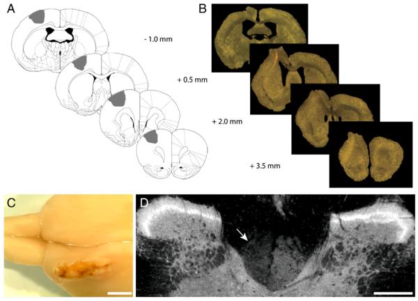
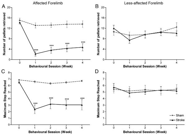
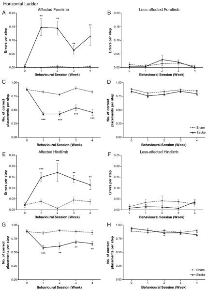
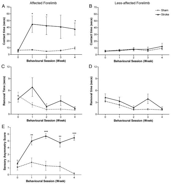
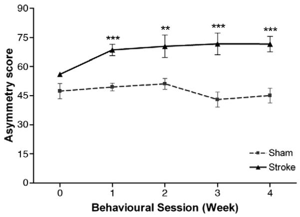
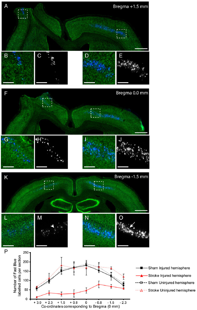
Despite recent advances, stroke remains a leading cause of neurological disability with the vast majority of victims being the elderly, who exhibit more severe neurological deficits and a reduced capacity to recover from these disabilities in comparison to young stroke survivors. The objective of the present study was to develop a model of focal ischemic stroke in aged rats using endothelin-1 (ET-1) to produce low mortality rates as well as reliable, robust sensorimotor deficits that resemble functional impairments associated with stroke in humans. Here, we studied the functional and histological outcome following unilateral ET-1 infusions into the sensorimotor cortex of aged rats (20-23 months old). This procedure resulted in low mortality rates (13.3%) and no loss in body weight one week following surgery. Functional assessment was performed using a number of reliable behavioural tests: staircase test (fine motor function), horizontal ladder (skilled locomotion), bilateral tactile stimulation test (somatosensory function) and cylinder test (postural weight support). Following ET-1 induced stroke, all tests demonstrated large and sustained sensorimotor deficits in both forelimb and hindlimb function that failed to improve over the 28-day testing period. In addition, histological assessment revealed a substantial loss of retrogradely labelled corticospinal neurons in the ipsilesional hemisphere following stroke. Our results establish a model for the use of aged rats in future preclinical studies, which will enhance assessment of the long-term benefit of potential neural repair and regenerative strategies.


Figures

Fig. 1
Fig. 1. Histological assessment of lesion
Representative illustrations of the lesion, demonstrating the AP co-ordinates of ET-1 administration relative to Bregma with the intended target area shaded in grey (A). The extent of ischemic damage through the brain can be seen in relation to the ET-1 injection sites in coronal sections viewed under a light microscope (B) and from a dorsal view of a rat brain 5 weeks post stroke in the left hemisphere (C). Immunostaining for PKCγ in transverse sections of the cervical spinal cord (C1) reveals the CST is intact on the right side and partially absent on the left side 5 weeks post stroke (D, indicated by arrow). Scale bars: 2 mm (C), 400 μm (D).
Fig. 2
Fig. 2. Staircase test
ET-1 stroke produced considerable impairments in fine motor function with rats unable to grasp many pellets successfully with their affected forepaw (A). Impairments were also observed in the rats' ability to reach and displace the sugar pellets (C), which also persisted throughout the entire testing period. A small but significant difference in the number of pellets retrieved was observed between sham and stroke rats using their less-affected forepaw (B) but no significant difference in maximum step reached (D) was observed between sham and stroke rats for their less-affected forepaw. Results are presented as mean±SEM and were analysed using RM ANCOVA and independent two-tailed t-tests. Significance between groups is denoted as: ***p<0.001. n=9 for sham and n=8 for stroke.
Fig. 3
Fig. 3. Horizontal ladder test
ET-1 stroke produced significant deficits in skilled locomotion, with animals exhibiting more footslip errors for both forelimb (A) and hindlimb (E) on the side contralateral to the stroke lesion compared to sham animals. Lesions also produced significant impairments in correct placement (score 6) with stroke animals unable to grasp the ladder rungs accurately with their affected forelimb (C) and hindlimb (G); these deficits persisted throughout the entire testing period. No significance between groups was observed in either errors per step or correct placement by the less-affected forelimb (B, D) or hindlimb (F, H). Results are presented as mean±SEM and were analysed using RM ANCOVA and independent two-tailed t-tests. Significance between groups is denoted as: **p<0.01; ***p<0.001. n=9 for sham and n=7 for stroke.
Fig. 4
Fig. 4. Bilateral tactile stimulation test
ET-1 stroke caused a significant impairment in somatosensory function. Lesions produced deficits in sensory awareness and sensorimotor function, with rats taking longer to contact (A) and remove (C) the adhesive patch from their affected paw compared to sham rats. No significant difference was observed to either contact (B) or remove (D) the adhesive patch from their less-affected paw. ET-1 lesions also caused a significant increase in somatosensory asymmetry (E). All deficits persisted throughout the entire testing period. Results are presented as mean±SEM and were analysed using RM ANCOVA and independent two-tailed t-tests. Significance between groups is denoted as: *p<0.05; **p<0.01; ***p<0.001. n=9 for sham, and n=8 for stroke.
Fig. 5
Fig. 5. Cylinder test
Normal rats display equal use in forelimb weight support with an asymmetry score of approximately 50. No significant difference between groups was found prior to surgery. After stroke, rats exhibited an increase in asymmetry indicating preferential use of the less-affected forelimb for support; this persisted throughout the entire testing period. Results are presented as mean±SEM and were analysed using RM ANCOVA and independent two-tailed t-tests. Significance between groups is denoted as: **p<0.01; ***p<0.001. n=9 for sham and n=8 for stroke.
Fig. 6
Fig. 6. Surviving corticospinal neurons after ET-1 stroke
Representative low (A, F, K) and high (B–E, G–J, L–O) power images of coronal sections through the brain immunostained for Nissl substance (Green). Sections reveal a substantial loss of retrogradely labelled CSNs (blue; black and white) in the injured (left) hemisphere in comparison to the uninjured (right) hemisphere 5 weeks after stroke. High power magnification highlights the striking difference between the severely atrophied somata (L, M, indicated by arrowhead) in the ipsilesional hemisphere and large healthy somata in the contralesional hemisphere (N, O, indicated by arrow). The distribution of CSNs in both hemispheres of sham and stroke rats revealed the extent of damage within the entire sensorimotor cortex after ET-1 induced stroke (P). Scale bars: 1 mm (A, F, K), 400 μm(B–E, G–J, L–O).

References

    1. Adams FS, Schwarting RK, Huston JP. Behavioral and neurochemical asymmetries following unilateral trephination of the rat skull: is this control operation always appropriate? Physiol. Behav. 1994;55:947–952. - PubMed
    1. Adkins DL, Voorhies AC, Jones TA. Behavioral and neuroplastic effects of focal endothelin-1 induced sensorimotor cortex lesions. Neuroscience. 2004;128:473–486. - PubMed
    1. Alloway KD, Hoffer ZS, Hoover JE. Quantitative comparisons of corticothalamic topography within the ventrobasal complex and the posterior nucleus of the rodent thalamus. Brain Res. 2003;968:54–68. - PubMed
    1. Badan I, Buchhold B, Hamm A, Gratz M, Walker LC, Platt D, Kessler C, Popa-Wagner A. Accelerated glial reactivity to stroke in aged rats correlates with reduced functional recovery. J. Cereb. Blood Flow Metab. 2003;23:845–854. - PubMed
    1. Barritt AW, Davies M, Marchand F, Hartley R, Grist J, Yip P, McMahon SB, Bradbury EJ. Chondroitinase ABC promotes sprouting of intact and injured spinal systems after spinal cord injury. J. Neurosci. 2006;26:10856–10867. - PMC - PubMed

Publication types

MeSH terms