. 2010 Apr;48(4):702-12.
doi: 10.1016/j.yjmcc.2009.10.032. Epub 2009 Nov 11.

Gene manipulated peritoneal cell patch repairs infarcted myocardium

Affiliations

Gene manipulated peritoneal cell patch repairs infarcted myocardium

Wei Huang et al. J Mol Cell Cardiol. 2010 Apr.

Abstract

A gene manipulated cell patch using a homologous peritoneum substrate was developed and applied after myocardial infarction to repair scarred myocardium. We genetically engineered male rat mesenchymal stem cells (MSC) using adenoviral transduction to over-express CXCR4/green fluorescent protein (GFP) (MSC(CXCR4)) or MSC(Null) or siRNA targeting CXCR4 (MSC(siRNA)). Gene expression was studied by real-time quantitative PCR (qPCR) and enzyme-linked immunosorbent assay (ELISA). Cells were cultured on excised peritoneum for 9 days. Two weeks after left anterior descending (LAD) coronary artery ligation in female hearts, the peritoneum patch was applied over the scarred myocardium, cell side down. Efficacy of engraftment was determined by presence of GFP positive cells. One month after cell implantation, echocardiography was performed and hearts were harvested for histological analysis. Left ventricle (LV) fibrosis, LV anterior wall thickness (AWT) and blood vessel density at the margins of the graft were measured. There was significant up-regulation of the chemokines in the MSC(CXCR4) group cultured under normoxic conditions when compared to the MSC(Null) group and a further increase was observed after exposure to hypoxia. One month after cell transplantation with the peritoneum patch, substantial numbers of GFP-positive cells were observed in and around the infarcted myocardium in MSC(CXCR4) group. LV AWT, LV fibrosis and LV function were significantly improved in the MSC(CXCR4) group as compared to these same variables in the MSC(Null) control. These salutary effects were absent in MSC(siRNA) group. The gene manipulated MSC-seeded peritoneum patch promotes tissue nutrition (angiogenesis), reduces myocardial remodeling, and enhances heart function after myocardial infarction.


Figures

Fig. 1
Fig. 1
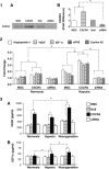
Efficiency of siRNA-mediated CXCR4 knockdown in MSC. Western blot shows the transfection efficiency of siRNA in MSC (1.1A). CXCR4 expression was knocked down by siRNA targeting CXCR4 gene (1.1B). Values mean ± SEM, n = 6 for each group. *p≤0.05 vs. MSC group. CXCR4, MSCCXCR4; Null, MSCNull; siRNA, MSCsiRNA; 1.2: Quantitative data of MSCCXCR4 for enhancing expression of chemokines. Angiopoietin 1, an angiogenic factor; VEGF, vascular endothelial growth factor; IGF-1α, insulin growth factor-1alpha; eIF4E, eukaryotic initiation factor 4E; cyclin A2, a cell-cycle-perpetuating. MSC, MSCNull; CXCR4, MSCCXCR4; siRNA, MSCsiRNA; *p≤0.05. All values expressed as mean ± SEM. n = 6 for each group. 1.3: ELISA results for release of VEGF (A) and IGF-1α protein (B) in cell lysate samples from ordinary MSC (MSC), Null (MSCNull), CXCR4 (MSCCXCR4), siRNA (MSCsiRNA) groups under normoxic, immediately after 8-h hypoxic, and 2-h re-oxygenation conditions. *p≤0.05. All values expressed as mean ± SEM. n = 6 for each group. Fig. 1.4: Schematic depiction of experimental design. The posterior abdominal peritoneum was clipped by two overlapping plastic-rings (A); Cut peritoneum with ring was shifted to cell culture dish and MSC were seeded on the peritoneum patch (B); MSC at 4, 7, and 9 days after seeding on the peritoneum patch in the dish (C). GFP-expressing monolayered MSC expanded to confluence on the peritoneum patch at day 9 and ready to use (D). The cell patch was grafted to the surface scared area of myocardium (E).
Fig. 1
Fig. 1
Efficiency of siRNA-mediated CXCR4 knockdown in MSC. Western blot shows the transfection efficiency of siRNA in MSC (1.1A). CXCR4 expression was knocked down by siRNA targeting CXCR4 gene (1.1B). Values mean ± SEM, n = 6 for each group. *p≤0.05 vs. MSC group. CXCR4, MSCCXCR4; Null, MSCNull; siRNA, MSCsiRNA; 1.2: Quantitative data of MSCCXCR4 for enhancing expression of chemokines. Angiopoietin 1, an angiogenic factor; VEGF, vascular endothelial growth factor; IGF-1α, insulin growth factor-1alpha; eIF4E, eukaryotic initiation factor 4E; cyclin A2, a cell-cycle-perpetuating. MSC, MSCNull; CXCR4, MSCCXCR4; siRNA, MSCsiRNA; *p≤0.05. All values expressed as mean ± SEM. n = 6 for each group. 1.3: ELISA results for release of VEGF (A) and IGF-1α protein (B) in cell lysate samples from ordinary MSC (MSC), Null (MSCNull), CXCR4 (MSCCXCR4), siRNA (MSCsiRNA) groups under normoxic, immediately after 8-h hypoxic, and 2-h re-oxygenation conditions. *p≤0.05. All values expressed as mean ± SEM. n = 6 for each group. Fig. 1.4: Schematic depiction of experimental design. The posterior abdominal peritoneum was clipped by two overlapping plastic-rings (A); Cut peritoneum with ring was shifted to cell culture dish and MSC were seeded on the peritoneum patch (B); MSC at 4, 7, and 9 days after seeding on the peritoneum patch in the dish (C). GFP-expressing monolayered MSC expanded to confluence on the peritoneum patch at day 9 and ready to use (D). The cell patch was grafted to the surface scared area of myocardium (E).
Fig. 2
Fig. 2
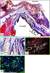
Characteristics of MSCCXCR4 in seeded peritoneal patch. Light microscopy showed that Masson's Trichome staining for characteristics of MSCCXCR4 in seeded peritoneal patch applied to the epicardium of the scared area at 4 weeks after cell patch implantation (A, B), (data in other groups not shown). Fluorescence microscopy demonstrated that newly formed blood vessels from infarcted areas (C) and inside of cell patch (D): In panel C, α-sarcomeric actin (red color) was used to identify myocytes, DAPI was used to identify all nuclei (blue color), and vessels derived from GFP positive MSC were identified by GFP antibody staining (green color). A = Original magnification × 100. B and C = Original magnification × 200; D = Original magnification × 400.
Fig. 3
Fig. 3
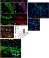
MSCCXCR4 migration and differentiation in situ at 4 weeks after patch graft. (A, B) shows immunofluorescent staining in CXCR4-P group (data in other groups not shown). Magnification × 100. (C) A cluster of enhanced GFP positive MSCCXCR4 originating from peritoneum patch migrated and localized in the infarct area (green fluorescence, arrow). (D) Same section as C except α-sarcomeric actin staining for cardiac myocytes (red fluorescence). (E) Same section as C and D except for co-localization of α-sarcomeric actin, EGFP and DAPI. (F) Enlarged picture from E (magnification × 400). C, D and E = Magnification × 200. (G) Quantitative data showed that the number of GFP positive MSC was significantly higher in CXCR4-P group than any other groups. MI + MSC-P, MI with MSCnull seeded patch treatment; MI + CXCR4-P, MI with MSCCXCR4 seeded patch treatment; MI + siRNA-P, MI with MSCsiRNA seeded patch treatment. All values were expressed as mean ± SEM. *p≤0.05; n = 6 for each group. (H) Some of the cells in CXCR4-P group were differentiated into cardiomyocytes (green color) which were confirmed by FISH analysis for Y-chromosome nuclei (arrow, red point). H = Magnification × 630.
Fig. 4
Fig. 4
Effects of MSCCXCR4 transplantation on angiogenesis in the infarct area. CXCR4-P group promotes vessel formation by significant expression of CD31 (green color; A, B, and C) and SMA (red color; D, E, and F) respectively in comparison with MSC-P and siRNA-P group, *p≤0.05 (magnification × 200). Quantitative analysis of capillary density (anti-CD31 antibody staining; G) and vascular density (anti α-smooth muscle actin antibody staining; H) was determined by various treatments. MI indicates myocardial infarction alone; MI + P, MI with patch treatment alone; MI + MSC-P, MI with MSCnull seeded patch treatment; MI + CXCR4-P, MI with MSCCXCR4 seeded patch treatment; MI + siRNA-P, MI with MSCsiRNA seeded patch treatment. All values expressed as mean ± SEM. *p≤0.05; n = 6 for each group.
Fig. 5
Fig. 5
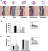
LV fibrosis and AWT in various treatment groups. At 4 weeks after cell patch treatment, left ventricular fibrosis and anterior wall thickness (AWT) were significantly attenuated and restoration of AWT was significantly increased in the MSC-P and CXCR4-P group as compared with other groups (A). AWT was thickest in CXCR4-P group (*p≤0.05). Quantitative data were shown for LV fibrosis (B) and AWT (C). The scale bars indicate 1.0 mm. MI indicates myocardial infarction alone; MI + P, MI with patch treatment alone; MI + MSC-P, MI with MSCnull seeded patch treatment; MI + CXCR4-P, MI with MSCCXCR4 seeded patch treatment; MI + siRNA-P, MI with MSCsiRNA seeded patch treatment. All values expressed as mean ± SEM. *p≤0.05; n = 12 for each group.
Fig. 6
Fig. 6
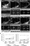
Cardiac function assessment by echocardiography at 4 weeks after cell patch transplantation (A). M-mode echocardiograms are shown at 1 day before, 1 week and 4 weeks after cell patch implantation in various treatment groups (B). Sham indicates open-chest state without LAD occlusion; MI, myocardial infarction alone; MI + P, MI with patch treatment alone; MI + MSC-P, MI with MSCNull seeded patch treatment; MI + CXCR4-P, MI with MSCCXCR4 seeded patch treatment; MI + siRNA-P, MI with MSCsiRNA seeded patch treatment. All values expressed as mean ± SEM. *p≤0.05 vs. MI, or MI + P, or MI + siRNA-P; #p≤0.05 vs. any other group; n = 12 for each group.

References

    1. Almsherqi ZA, McLachlan CS, Slocinska MB, Sluse FE, Navet R, Kocherginsky N, et al. Reduced cardiac output is associated with decreased mitochondrial efficiency in the non-ischemic ventricular wall of the acute myocardial-infarcted dog. Cell Res. 2006;16:297–305. - PubMed
    1. Taheri SA, Ashraf H, Merhige M, Miletich RS, Satchidanand S, Malik C, et al. Myoangiogenesis after cell patch cardiomyoplasty and omentopexy in a patient with ischemic cardiomyopathy. Tex Heart Inst J. 2005;32:598–601. - PMC - PubMed
    1. O'Leary DP. Use of the greater omentum in colorectal surgery. Dis Colon Rectum. 1999;42:533–9. - PubMed
    1. O'Shaughnessy L. Surgical treatment of cardiac ischemia. Lancet. 1937;232:185–94.
    1. Suzuki R, Hattori F, Itabashi Y, Yoshioka M, Yuasa S, Manabe-Kawaguchi H, et al. Omentopexy enhances graft function in myocardial cell sheet transplantation. Biochem Biophys Res Commun. 2009;387:353–9. - PubMed

Publication types

MeSH terms