

The neuronal correlates of intranasal trigeminal function-an ALE meta-analysis of human functional brain imaging data
- PMID: 19913573
- PMCID: PMC2822005
- DOI: 10.1016/j.brainresrev.2009.11.001
The neuronal correlates of intranasal trigeminal function-an ALE meta-analysis of human functional brain imaging data
Abstract
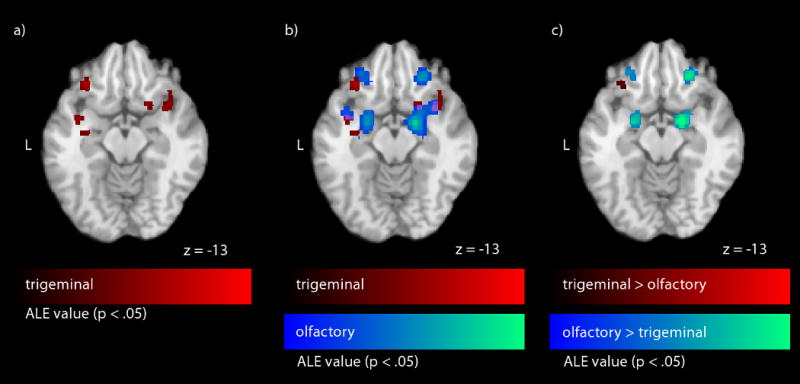
Almost every odor we encounter in daily life has the capacity to produce a trigeminal sensation. Surprisingly, few functional imaging studies exploring human neuronal correlates of intranasal trigeminal function exist, and results are to some degree inconsistent. We utilized activation likelihood estimation (ALE), a quantitative voxel-based meta-analysis tool, to analyze functional imaging data (fMRI/PET) following intranasal trigeminal stimulation with carbon dioxide (CO(2)), a stimulus known to exclusively activate the trigeminal system. Meta-analysis tools are able to identify activations common across studies, thereby enabling activation mapping with higher certainty. Activation foci of nine studies utilizing trigeminal stimulation were included in the meta-analysis. We found significant ALE scores, thus indicating consistent activation across studies, in the brainstem, ventrolateral posterior thalamic nucleus, anterior cingulate cortex, insula, precentral gyrus, as well as in primary and secondary somatosensory cortices-a network known for the processing of intranasal nociceptive stimuli. Significant ALE values were also observed in the piriform cortex, insula, and the orbitofrontal cortex, areas known to process chemosensory stimuli, and in association cortices. Additionally, the trigeminal ALE statistics were directly compared with ALE statistics originating from olfactory stimulation, demonstrating considerable overlap in activation. In conclusion, the results of this meta-analysis map the human neuronal correlates of intranasal trigeminal stimulation with high statistical certainty and demonstrate that the cortical areas recruited during the processing of intranasal CO(2) stimuli include those outside traditional trigeminal areas. Moreover, through illustrations of the considerable overlap between brain areas that process trigeminal and olfactory information; these results demonstrate the interconnectivity of flavor processing.
Figures


References
-
- Albrecht J, Kopietz R, Kleemann AM, Schöpf V, Fesl G, Anzinger A, Schreder T, Kobal G, Wiesmann M. Brain activation of olfactory and trigeminal cortical areas is independent from perceptual strength – a functional magnetic resonance imaging study using nicotine as chemosensory stimulus. Chem Senses. 2007;32:A124.
-
- Albrecht J, Kopietz R, Linn J, Sakar V, Anzinger A, Schreder T, Pollatos O, Bruckmann H, Kobal G, Wiesmann M. Activation of olfactory and trigeminal cortical areas following stimulation of the nasal mucosa with low concentrations of S(-)-nicotine vapor – an fMRI study on chemosensory perception. Hum Brain Mapp. 2009;30:699–710. - PMC - PubMed
-
- Alvaro RE, Weintraub Z, Kwiatkowski K, Cates DB, Rigatto H. A respiratory sensory reflex in response to CO2 inhibits breathing in preterm infants. J Appl Physiol. 1992;73:1558–63. - PubMed
-
- Anton F, Peppel P, Euchner I, Handwerker HO. Controlled noxious chemical stimulation: responses of rat trigeminal brainstem neurones to CO2 pulses applied to the nasal mucosa. Neurosci Lett. 1991;123:208–11. - PubMed
-
- Bensafi M, Sobel N, Khan RM. Hedonic-specific activity in piriform cortex during odor imagery mimics that during odor perception. J Neurophysiol. 2007;98:3254–62. - PubMed
Publication types
MeSH terms
Grants and funding
LinkOut - more resources
Full Text Sources
