

Mechanics of the F-actin cytoskeleton
- PMID: 19913792
- PMCID: PMC2813332
- DOI: 10.1016/j.jbiomech.2009.09.003
Mechanics of the F-actin cytoskeleton
Abstract
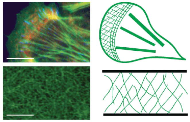
Dynamic regulation of the filamentous actin (F-actin) cytoskeleton is critical to numerous physical cellular processes, including cell adhesion, migration and division. Each of these processes require precise regulation of cell shape and mechanical force generation which, to a large degree, is regulated by the dynamic mechanical behaviors of a diverse assortment of F-actin networks and bundles. In this review, we review the current understanding of the mechanics of F-actin networks and identify areas of further research needed to establish physical models. We first review our understanding of the mechanical behaviors of F-actin networks reconstituted in vitro, with a focus on the nonlinear mechanical response and behavior of "active" F-actin networks. We then explore the types of mechanical response measured of cytoskeletal F-actin networks and bundles formed in living cells and identify how these measurements correspond to those performed on reconstituted F-actin networks formed in vitro. Together, these approaches identify the challenges and opportunities in the study of living cytoskeletal matter.
Copyright 2009 Elsevier Ltd. All rights reserved.
Figures

References
-
- Pollard TD. Cytoskeletal functions of cytoplasmic contractile proteins. J Supramol Struct. 1976;5(3):317–34. - PubMed
-
- Clarke M, Spudich JA. Nonmuscle contractile proteins: the role of actin and myosin in cell motility and shape determination. Annu Rev Biochem. 1977;46:797–822. - PubMed
-
- Stossel TP. Contractile proteins in cell structure and function. Annu Rev Med. 1978;29:427–57. - PubMed
-
- Janmey PA, et al. Resemblance of actin-binding protein/actin gels to covalently crosslinked networks. Nature. 1990;345(6270):89–92. - PubMed
Publication types
MeSH terms
Substances
Grants and funding
LinkOut - more resources
Full Text Sources
