. 2009 Nov 13;139(4):780-90.
doi: 10.1016/j.cell.2009.10.010.

Structural convergence between Cryo-EM and NMR reveals intersubunit interactions critical for HIV-1 capsid function

Affiliations

Structural convergence between Cryo-EM and NMR reveals intersubunit interactions critical for HIV-1 capsid function

In-Ja L Byeon et al. Cell. .

Abstract

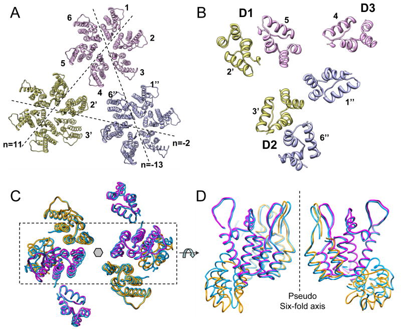
Mature HIV-1 particles contain conical-shaped capsids that enclose the viral RNA genome and perform essential functions in the virus life cycle. Previous structural analysis of two- and three-dimensional arrays of the capsid protein (CA) hexamer revealed three interfaces. Here, we present a cryoEM study of a tubular assembly of CA and a high-resolution NMR structure of the CA C-terminal domain (CTD) dimer. In the solution dimer structure, the monomers exhibit different relative orientations compared to previous X-ray structures. The solution structure fits well into the EM density map, suggesting that the dimer interface is retained in the assembled CA. We also identified a CTD-CTD interface at the local three-fold axis in the cryoEM map and confirmed its functional importance by mutagenesis. In the tubular assembly, CA intermolecular interfaces vary slightly, accommodating the asymmetry present in tubes. This provides the necessary plasticity to allow for controlled virus capsid dis/assembly.


Figures

Figure 1
Figure 1
CryoEM and 3D reconstruction of full-length HIV-1 CA tubular assemblies. CryoEM micrograph of recombinant wild-type (WT) HIV-1 CA (A) and HIV-1 CA A92E (B). Scale bars, 100 nm. (C&D) An enlarged A92E tube image and its computed Fourier transforms. Scale bar, 50 nm. The arrow points to the layerline at 20Å resolution. (E–G) 3D reconstruction of the A92E CA tubes from the (−13, 11) helical family. The density map of the A92E CA tube is displayed as three orthogonal slices: parallel to the tube axis and close to the surface (E), perpendicular to the tube axis (F), and parallel to and through the tube axis (G). Scale bars, 10 nm.
Figure 2
Figure 2
Structure of the HIV-1 CA helical assembly and domain docking. (A) Surface rendering of the reconstructed tubular structure contoured at 2.3σ (blue) and 1.3σ (gold) enclosing 65% and 100% volume, respectively. Dashed lines connect hexamers in the three distinct helical arrangements denoted as n=−2, 11, and −13 helices; n is the Bessel order. (B–D) Docking of NTD and CTD domain models independently into the tubular density map (contour enclosing 90% volume). Shown are a view from the tube surface (B) and slab views to show the NTD region (C) and the CTD region (D). Three docked hexamers are displayed. A local threefold axis is marked by *.
Figure 3
Figure 3
Asymmetric arrangement of CA hexamers in the tubular assembly. (A) The pseudo-atomic domain docking model of a trimer of NTD hexamers shown with the same orientation as in Figure 2B–D. Dashed lines denote the n=−2, 11, and −13 helices. The six monomers in each hexamer are numbered 1–6. (B) Domain docking model of a trimer of CTD dimers between adjacent hexamers, denoted as D1, D2, and D3, at a local three-fold axis. (C) Domain docked monomers (only 2, 3, 5 and 6 are shown), superimposed on to the corresponding monomers (blue) in the six-fold symmetric hexamer docked model. Monomer 2 and 5 (magenta), as well as 3 and 6 (orange), are related by two-fold symmetry. (D) A 90° rotated view of the boxed region in (C) illustrates that the NTDs and CTDs of monomers 3 and 6 (orange) are offset from six-fold symmetry, whereas monomers 2 and 4 (magenta) exhibit little shift.
Figure 4
Figure 4
Spectral comparison between full-length CA, the NTD CA, and the monomer and dimer states of CTD CA. (A) Superposition of the 900 MHz 1H,15N HSQC spectra of full-length (blue), NTD (green), and CTD (red) domains. Selected backbone amide and Trp indole HNε resonances of full-length CA are labeled by residue name and number in the non-crowded regions of the spectrum. The protein concentration in all samples was 0.15mM. The folded Gly106 peak is marked by *. (B) Superposition of the 900 MHz 1H,15N HSQC spectra of CTD at two concentrations, 2mM (red) and 0.0124mM (cyan). Resonances for the dimer state (red) are labeled with assignments, and those only present in the dimer are circled.
Figure 5
Figure 5
NMR solution structure of the CTD dimer. (A) Backbone (N, Cα, C′) superpositions of the final 30 conformer ensemble. Monomer #1 is colored pink, with helix 9 in magenta, and monomer #2 is shown in grey, with helix 9 in cyan. (B) Stereoview of a ribbon representation for an individual structure of the ensemble using the color scheme of (A). Seven amino acids that are involved in extensive hydrophobic interactions at the dimer interface are shown in stick representation in gold (monomer #1) and blue (monomer #2), respectively, and labeled with residue name and number. (C) Local structure surrounding Tyr145. The color scheme is identical to (B). (D) Intermolecular NOEs involving Tyr145 detected in the 3D 13C/15N-filtered, 13C-edited NOESY spectrum. (E) Comparison of core-associated CA (black), infectivity (green), and RT activity (purple) for WT, Y145A, and Y145F HIV-1 particles.
Figure 6
Figure 6
Comparison between the present NMR solution structure of the CTD-CA dimer and previously reported CTD dimer structures from X-ray crystallography and cryoEM. (A) Backbone atomic superpositions of the NMR structure (pink) and the crystal structures 1BAJ (blue), 1A8O (purple) and 2BUO (red orange) and the pseudo-atomic EM structure from 2D crystals, 3DIK (green). The best-fit superposition was carried out for one monomer unit to highlight the difference in subunit orientations in the dimers. (B) Superpositions of the central regions (residues 181–187) of helices 9 in the CTD dimer interfaces in ribbon representation: the top view highlights the differences in the shapes formed by these helices and the bottom views are rotated by −90° around the y-axis to emphasize the crossing angles. The color scheme is identical to (A). (C) Present cryoEM pseudo-atomic structure of capsid hexamers in tubes modeled using rigid body docking of an NTD monomer and the NMR CTD dimer into the EM map. The NTD is shown in yellow ribbon representation and the CTD is in magenta. (D) Superposition of the CTD dimer structures at the trimer interface of the current cryoEM model (magenta) and the 2D crystal model (pale green).
Figure 7
Figure 7
Inter-hexamer interactions at the local three-fold axis mediated by the CTD dimers. (A) Surface rendering of the density map (contoured with 2.0σ) displaying three neighboring hexamers. Local two-fold, three-fold, and six-fold symmetry axes are indicated by ellipses, a triangles, and hexagons, respectively. (B) Detailed view of the three-fold axis illustrating the interactions at the interface. Side chains of K203 and P207 in H10, and E213, T216, and Q219 in H11 are shown in ball and stick representation. Mutations of these residues are known to affect in vitro assembly and capsid stability. Two pairs of spatially close residues, P207/T216 and K203/Q219, were tested by cysteine cross-linking. (C) Virus infectivity of WT and the K203C/Q219C and P207C/T216C CA mutants. (D) Intermolecular cross-linking studies of HIV-1 particles prepared from cultured cells. Lanes 1&2, WT; lanes 3&4, K203C/Q219C; lanes 5&6, P207C/T216C; lanes 7, 8, 9, reduced (2-mercaptoethanol-treated) WT, K203C/Q219C, and P207C/T216C samples, respectively. (E) Comparison of core stability (black) and infectivity (green) of WT and the E213A and E213Q mutant particles. (F) Kinetics of HIV-1 uncoating of purified WT, E213A, and E213Q cores in vitro.

Similar articles

Cited by

References

    1. Aiken C. Cell-free assays for HIV-1 uncoating. Methods Mol Biol. 2009;485:41–53. - PMC - PubMed
    1. Alcaraz LA, del Alamo M, Barrera FN, Mateu MG, Neira JL. Flexibility in HIV-1 assembly subunits: solution structure of the monomeric C-terminal domain of the capsid protein. Biophys J. 2007;93:1264–1276. - PMC - PubMed
    1. Bax A, Grzesiek S. Methodological advances in protein NMR. Acc Chem Res. 1993;26:131–138.
    1. Berthet-Colominas C, Monaco S, Novelli A, Sibai G, Mallet F, Cusack S. Head-to-tail dimers and interdomain flexibility revealed by the crystal structure of HIV-1 capsid protein (p24) complexed with a monoclonal antibody Fab. Embo J. 1999;18:1124–1136. - PMC - PubMed
    1. Bosco DA, Kern D. Catalysis and binding of cyclophilin A with different HIV-1 capsid constructs. Biochemistry. 2004;43:6110–6119. - PubMed

Publication types

Associated data

LinkOut - more resources