. 2009 Nov 12;64(3):328-40.
doi: 10.1016/j.neuron.2009.08.036.

A functional mouse retroposed gene Rps23r1 reduces Alzheimer's beta-amyloid levels and tau phosphorylation

Affiliations

A functional mouse retroposed gene Rps23r1 reduces Alzheimer's beta-amyloid levels and tau phosphorylation

Yun-wu Zhang et al. Neuron. .

Abstract

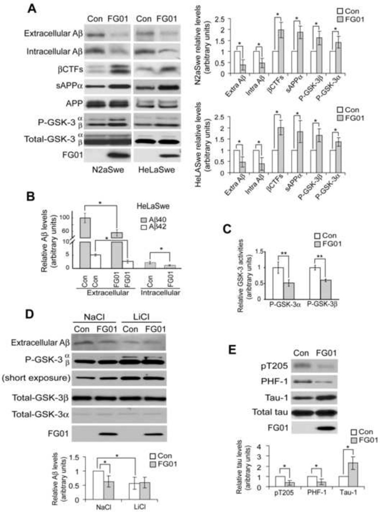
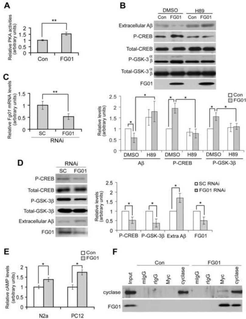
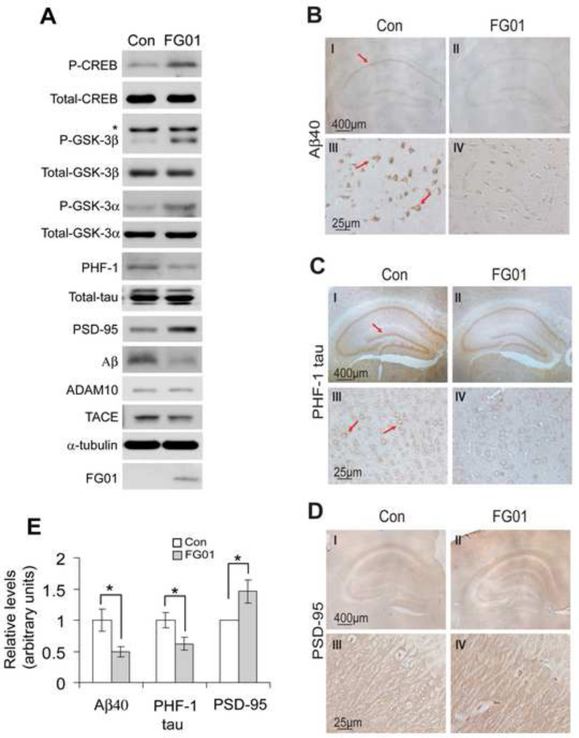
Senile plaques consisting of beta-amyloid (Abeta) and neurofibrillary tangles composed of hyperphosphorylated tau are major pathological hallmarks of Alzheimer's disease (AD). Elucidation of factors that modulate Abeta generation and tau hyperphosphorylation is crucial for AD intervention. Here, we identify a mouse gene Rps23r1 that originated through retroposition of ribosomal protein S23. We demonstrate that RPS23R1 protein reduces the levels of Abeta and tau phosphorylation by interacting with adenylate cyclases to activate cAMP/PKA and thus inhibit GSK-3 activity. The function of Rps23r1 is demonstrated in cells of various species including human, and in transgenic mice overexpressing RPS23R1. Furthermore, the AD-like pathologies of triple transgenic AD mice were improved and levels of synaptic maker proteins increased after crossing them with Rps23r1 transgenic mice. Our studies reveal a new target/pathway for regulating AD pathologies and uncover a retrogene and its role in regulating protein kinase pathways.


Figures

Figure 1
Figure 1. Genetic Screening Using Random Homozygous Gene Perturbation (RHGP)
(A) The new RHGP gene search vector has a tetracycline-regulated element (TRE) regulated CMV promoter, which drives expression of the puromycin N-acetyl-tranferase gene (pac), a plasmid replication origin and a chloramphenicol resistance marker (Ori-CAT), and a LoxP site in both the 5’LTR (not shown) and the 3’LTR. In addition, there is a Cre recombinase gene (Cre) between the 5’LTR and the 3’LTR. (B) The initial provirus randomly inserted into chromosomes of mammalian cells upon retroviral infection. (C) The final integrated provirus after the expression of the Cre recombinase in the initial provirus, which mediates DNA recombination at the loxP sites. (D) Strategy for screening for Aβ-reducing genes in N2aSwe cells with RHGP vector integration.
Figure 2
Figure 2. Identification of the FG01 Cell Clone
(A) RHGP libraries of N2aSwe cells were live-immunostained for cell surface βCTF using an Aβ N-terminal specific antibody (FCA18) (Barelli et al., 1997) and screened by multiple rounds of FACS sorting. Less than 0.01% of cells showing βCTF accumulation after first round of FACS sorting were enriched up to 75% following another two rounds of FACS sorting. Y axis, cell number; X axis, fluorescence intensity. (B) Parental N2aSwe cells (I and III) and one cell clone derived from FACS sorting, FG01 (II and IV), were live-immunostained to visualize surface APP βCTF (red, I and II). Cells were also double immunostained with FITC-VVA (Vicia Vilosa Agglutinin, Vector Laboratories) to stain total surface glycoproteins (III and IV). (C) Parental N2aSwe and FG01 cells were treated with or without 2 µg/ml doxycycline (DOX) for 72 hrs. Equal amounts of cell lysates were subjected to SDS-PAGE and Western blot to detect full length APP and βCTFs. Secreted Aβ was immunoprecipitated from conditioned media and analyzed by Western blot. Protein levels in FG01 cells were quantified by densitometry and normalized to those of controls for comparison (set as one arbitrary unit). (D) Parental N2aSwe and FG01 cells were treated with 2 µg/ml doxycycline (+) or DMSO (−) for 72 hrs before RNA was isolated for real-time reverse transcription-PCR to quantify Fg01 expression. The level of Fg01 in N2aSwe cells treated with DMSO was used as normalization controls (set as one arbitrary unit). *P<0.05. P values were calculated using two-tailed Student’s t-test (n = 3). Error bars, SEM.
Figure 3
Figure 3. The Fg01 Gene Originated Through Retroposition of the Mouse Rps23 mRNA
(A) Sequence alignment of the reverse and complementary (RC) sequence of mouse Rps23 (mRps23) mRNA with Fg01. Small letters indicate intron sequence (in green) or untranslated regions of the exon (in red). Capital letters indicate protein-encoding sequence (in blue). Additional gene parts of Fg01 recruited from integrated chromosomal sites were indicated by lines (for introns) and boxes (for exons) (not drawn to proportion). The colors red, green and blue indicate exons, introns, and protein-encoding regions, respectively. *: nonconserved nucleotide residues. (B) Phylogenetic relationships among Fg01 and human, rat and mouse Rps23 genes (hRps23, rRps23, and mRps23) based on their sequence identity to mRps23 within the homologous region.
Figure 4
Figure 4. FG01 is a Type Ib Transmembrane Protein
(A) Scheme of the FG01 construct used in this study (not drawn to proportion), with a Myc tag at the N-terminus and a His6 tag at the C-terminus. FG01 has a predicted single trans-membrane domain (TM) near its C-terminus. (B) The Myc-FG01-His6 vector or a pcDNA control was transiently transfected into N2a cells. Cell lysates were subjected to Western blot (WB) with antibodies against Myc or His6. (C) FG01, APP and SMAD3 plasmids (all Myc-tagged) were individually transfected into N2a cells. After fractionation of membrane and cytosol, equal volumes of samples from both fractions were subjected to SDS-PAGE and Western analysis with a Myc antibody. *: non-specific band. (D) After FG01 transfection, N2a cells were biotinylated and biotin-labeled membrane proteins were affinity precipitated (AP) with streptivadin and immunoblotted with a Myc antibody. (E) After transfection with the Myc-FG01-His6 construct, N2a cells were either live-immunostained or permeabilized and immunostained with Myc or His6 antibody. Cells were then fixed, permeabilized, incubated with Alexa Fluor 488-conjugated secondary antibody and DAPI, and examined by immunofluorescence microscopy. Red arrows indicate membrane staining of FG01 in live cells. (F) Equal protein lysates from mouse cortex and hippocampus (hippo) were incubated with an FG01 antibody (+) or rabbit IgG (−). After immunoprecipitation, samples were subjected to SDS-PAGE and Western blot analysis with the FG01 antibody. (G) An anti-sense probe of Fg01 and the corresponding sense probe (as control) were used for in situ hybridization in brain sections from a two-month-old C57Bl6 mouse. Red arrows indicate Fg01 expression.
Figure 5
Figure 5. FG01 Reduces Aβ Levels, GSK-3 Activity and tau Phosphorylation
(A) FG01 or control vector (Con) were transfected into mouse N2aSwe or human HeLaSwe cells. Aβ in conditioned media (secreted or extracellular) and cell lysates (intracellular) was immunoprecipitated and Western blotted with the Aβ antibody 6E10. sAPPα in conditioned media was immunoblotted with 6E10. Cell lysates were immunoblotted with antibodies against APP/βCTF (6E10), phosphorylated GSK-3α/β, total GSK3α/β, and Myc-FG01 (9E10). (B) ELISA quantification of Aβ40 and Aβ42 levels in conditioned media and lysates of HelaSwe cells with FG01 overexpression. Results were normalized to that of Aβ40 in conditioned media (set as 100). Aβ42 in cell lysates was below detection level and not shown. (C) GSK-3α and GSK-3β in lysates from N2aSwe cells transfected with control or FG01 cDNA were immunoprecipitated with respective antibodies and assayed for in vitro activity. Results were normalized to those of controls (set as one arbitrary unit). (D) N2aSwe cells were first transfected with FG01 or control vector (Con). After equal splitting, cells were treated with 5 mM LiCl or NaCl (as control) for 4 hrs before collection. Conditioned media were assayed for Aβ. Cell lysates were analyzed for total and phosphorylated GSK-3α/β and for FG01. (E) N2a cells were transfected with human tau, equally split, and transfected with FG01 or control vector (Con). The levels of phosphorylated tau including threonine 205 (pT205) and PHF-1, unphopshorylated tau (Tau-1), total tau, and FG01 were analyzed. In some experiments, protein levels were quantified by densitometry and normalized to those of controls for comparison (set as one arbitrary unit). All error bars indicate SEM. *P<0.05, **P<0.01. P values were calculated using two-tailed Student’s t-test (n = 3).
Figure 6
Figure 6. FG01 Interacts with Adenylate Cyclases, Upregulates cAMP Levels and Activates PKA to Reduce GSK-3 Activity and Aβ Levels
(A) Cells transfected with FG01 or control vector (Con) were analyzed for in vitro PKA activity. Results were normalized to control values (set as one arbitrary unit). (B) After transfection with FG01 or control vectors (Con) and equal splitting, cells were treated with DMSO (control) or the PKA inhibitor H89. Conditioned media were analyzed for Aβ and cell lysates were analyzed for phosphorylated and total CREB, phosphorylated and total GSK-3, and FG01 levels. (C) N2aSwe cells were transfected with Fg01-specific RNAi or a scrambled RNAi (SC). Total RNA was then extracted and subjected to RT-PCR. The level of Fg01 relative to that of β-actin was analyzed and normalized to that from scrambled RNAi-transfected cells (set as one arbitrary unit). (D) After RNAi of Fg01 expression, conditioned media from N2aSwe cells were analyzed for Aβ, and cell lysates were analyzed for endogenous FG01 and phosphorylated/total CREB and GSK-3. (E) Mouse N2a and rat PC12 cells were transfected with FG01 or control vectors, and cell lysates were assayed for cAMP levels. Data were normalized to control values (as one arbitrary unit). (F) Cells transfected with FG01 or control vectors were lysed in 1% CHAPSO or 1% NP40 buffer. Lysates were incubated with mouse IgG (mIgG), rabbit IgG (rIgG), Myc antibody or adenylate cyclase antibody. Immunoprecipitated proteins were subjected to SDS-PAGE and Western blot analysis with adenylate cyclase or Myc (for FG01) anatibodies. In some experiments, protein levels were quantified by densitometry and normalized to those of controls for comparison (set as one arbitrary unit). All error bars indicate SEM. *P<0.05, **P<0.01. P values were calculated using two-tailed Student’s t-test (n = 3).
Figure 7
Figure 7. FG01 Overexpression Increases PKA Activity and Synapse number and Reduces GSK-3β Activity, tau Phosphorylation, and Aβ Levels in Brains of 3XTg AD Mice
(A) Brains from FG01 mice and littermate controls (Con) on a 3XTg background at 11 months of age were dissected. One half of the brain was lysed and analyzed for the levels of phosphorylated/total CREB, phosphorylated/total GSK-3, PSD-95, ADAM10, TACE, α-tubulin, and phosphorylated (PHF-1) and total tau forms by direct Western blot. Aβ and Myc-FG01 were detected by immunoprecipitation-Western blot using Aβ antibody (6E10) and Myc antibody, respectively. *: non-specific band found specifically in 3XTg mouse brains. (B) The other half brain from Con (I and III) and FG01 (II and IV) mice was analyzed by immunohistochemistry for Aβ using an Aβ40-specific antibody. III and IV are higher magnifications of cortical regions from I and II, respectively. Red arrows indicate positive immunoreactivity. (C) Immunohistochemistry for phosphorylated tau (PHF-1 tau) was analyzed the same as in (B). Red arrows indicate positive immunoreactivity. (D) Immunohistochemistry for PSD-95 was analyzed the same as in (B), except that III and IV are higher magnifications of hippocampal regions from I and II, respectively. (E) Immunostained neurons (>400) in (B) and (C) were counted from 5 randomly selected cortical regions. Ratios of Aβ40-positive and PHF-1 tau-positive neurons to total neurons were determined and normalized to those of control (Con) for comparison. The optical density (darkness) of PSD-95 staining in 5 randomly selected hippocampal regions were analyzed by the Photoshop software for comparison. *P<0.05. P values were calculated using two-tailed Student’s t-test (n = 4). Error bars, SEM.

Similar articles

Cited by

References

    1. Ancolio K, Dumanchin C, Barelli H, Warter JM, Brice A, Campion D, Frebourg T, Checler F. Unusual phenotypic alteration of beta amyloid precursor protein (betaAPP) maturation by a new Val-715 --> Met betaAPP-770 mutation responsible for probable early-onset Alzheimer’s disease. Proc. Natl. Acad. Sci. USA. 1999;96:4119–4124. - PMC - PubMed
    1. Babushok DV, Ostertag EM, Kazazian HH., Jr. Current topics in genome evolution: molecular mechanisms of new gene formation. Cell. Mol. Life. Sci. 2007;64:542–554. - PMC - PubMed
    1. Barelli H, Lebeau A, Vizzavona J, Delaere P, Chevallier N, Drouot C, Marambaud P, Ancolio K, Buxbaum JD, Khorkova O, et al. Characterization of new polyclonal antibodies specific for 40 and 42 amino acid-long amyloid beta peptides: their use to examine the cell biology of presenilins and the immunohistochemistry of sporadic Alzheimer’s disease and cerebral amyloid angiopathy cases. Mol. Med. 1997;3:695–707. - PMC - PubMed
    1. Buee L, Bussiere T, Buee-Scherrer V, Delacourte A, Hof PR. Tau protein isoforms, phosphorylation and role in neurodegenerative disorders. Brain Res. Brain Res. Rev. 2000;33:95–130. - PubMed
    1. Buxbaum JD, Ruefli AA, Parker CA, Cypess AM, Greengard P. Calcium regulates processing of the Alzheimer amyloid protein precursor in a protein kinase C-independent manner. Proc. Natl. Acad. Sci. USA. 1994;91:4489–4493. - PMC - PubMed

Publication types

MeSH terms