. 2009 Nov 12;64(3):341-54.
doi: 10.1016/j.neuron.2009.10.023.

Math1 is essential for the development of hindbrain neurons critical for perinatal breathing

Affiliations

Math1 is essential for the development of hindbrain neurons critical for perinatal breathing

Matthew F Rose et al. Neuron. .

Abstract

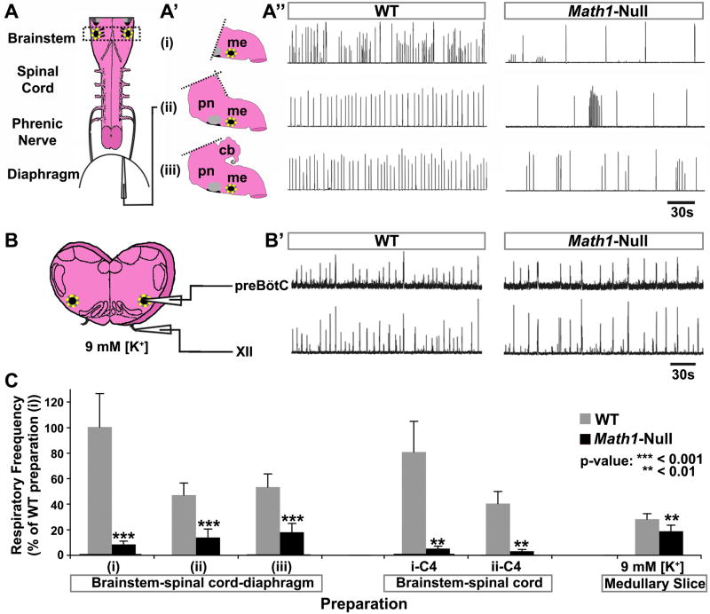
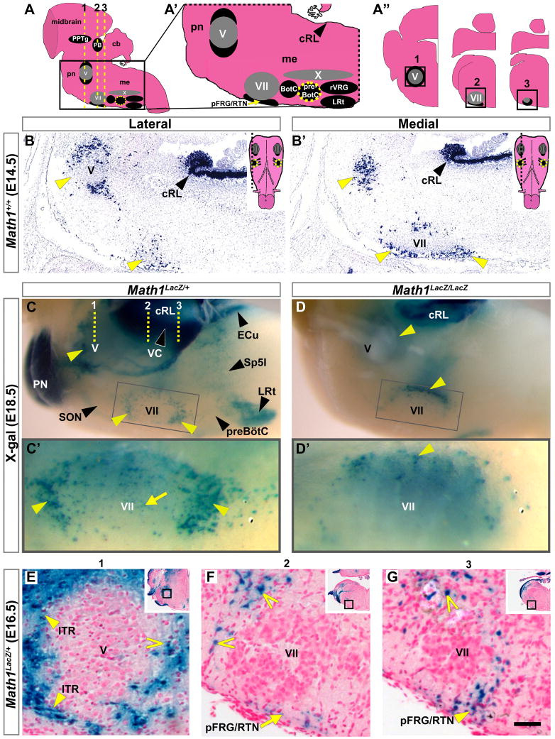
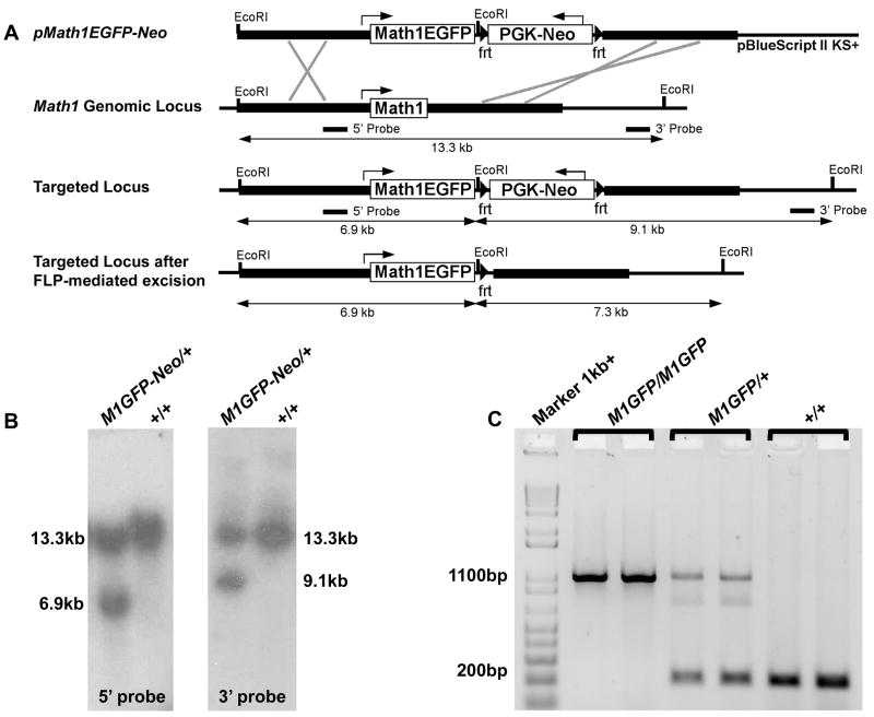
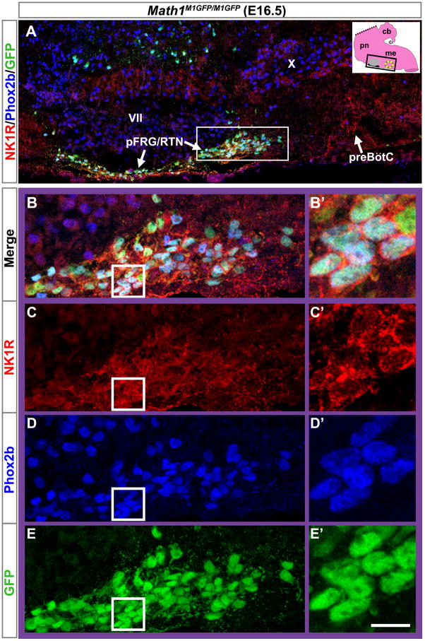
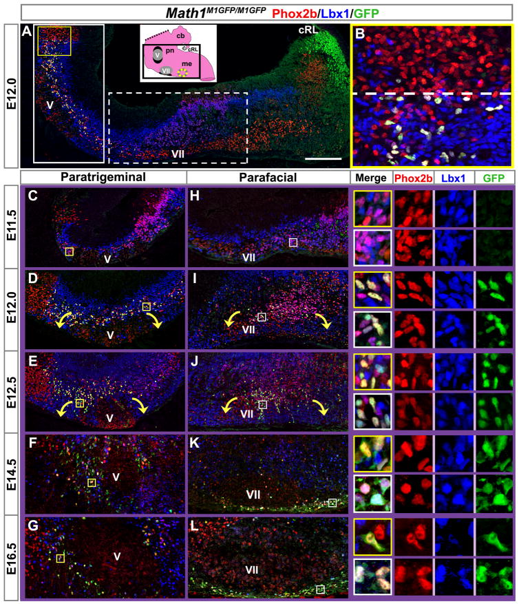
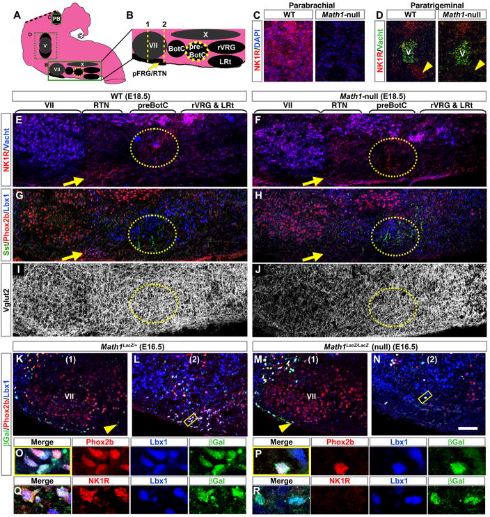
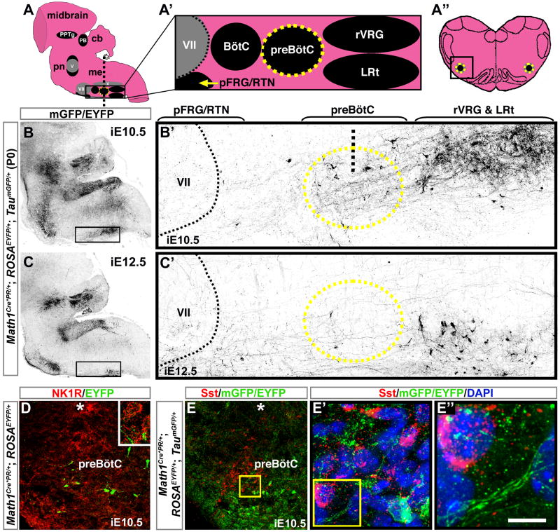
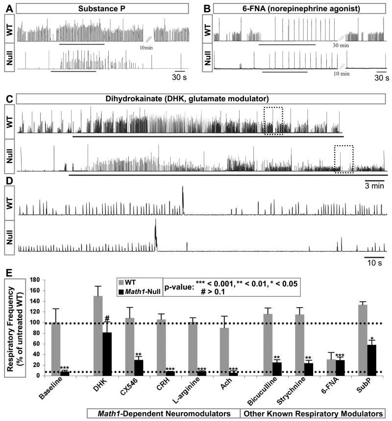
Mice lacking the proneural transcription factor Math1 (Atoh1) lack multiple neurons of the proprioceptive and arousal systems and die shortly after birth from an apparent inability to initiate respiration. We sought to determine whether Math1 was necessary for the development of hindbrain nuclei involved in respiratory rhythm generation, such as the parafacial respiratory group/retrotrapezoid nucleus (pFRG/RTN), defects in which are associated with congenital central hypoventilation syndrome (CCHS). We generated a Math1-GFP fusion allele to trace the development of Math1-expressing pFRG/RTN and paratrigeminal neurons and found that loss of Math1 did indeed disrupt their migration and differentiation. We also identified Math1-dependent neurons and their projections near the pre-Bötzinger complex, a structure critical for respiratory rhythmogenesis, and found that glutamatergic modulation reestablished a rhythm in the absence of Math1. This study identifies Math1-dependent neurons that are critical for perinatal breathing that may link proprioception and arousal with respiration.


Figures

Figure 1
Figure 1. Math1-Null Mice Die after Birth because of Too-Slow Central Respiratory Rhythm
(A) Schematic of the standard brainstem-spinal cord-diaphragm (BSD) preparation used for respiratory physiology (includes only the medullary portion of the brainstem). (A′) Schematics depicting three variations of the BSD preparation in which different portions of the brainstem were included: (i) medulla only, (ii) pons and medulla, and (iii) cerebellum, pons, and medulla. (A″) Rectified and integrated suction electrode recordings from the diaphragm muscle EMG in the three BSD preparations from E18.5 WT mice (left column) and Math1-null mice (right column). The rhythm was significantly slower and more irregular in all Math1-null preparations. Yellow dotted circle on each schematic indicates the preBötC, while the black region ventral to the facial nucleus (gray circle) represents the pFRG/RTN. (B) Schematic of medullary slice preparation containing the preBötC (from the region indicated by the dashed rectangle in (A)). (B′) Rectified and integrated suction electrode recordings from the preBötC (top) and XII motoneuron pool (bottom) of WT (left) and Math1-null (right) slice preparations (in 9mM [K+]). The frequency was again slower in Math1-null preparations but was more stable than in the BSD preparations. (C) Population data of respiratory frequency from WT (gray) and Math1-null (black) mice for each BSD preparation (i–iii) depicted in (A), for recordings directly from C4 in these preparations after removal of the diaphragm (i-C4: medulla only, ii-C4: pons and medulla), and for the medullary slice preparations. Data was normalized to that of WT preparation (i) for ease of comparison. P-values indicate a significant difference between Math1-null and WT tissue within each preparation. See Table S1 for frequency, sample size, and statistical analyses. Abbreviations: cerebellum (cb), hypoglossal motor nucleus (XII), medulla (me), pons (pn), pre-Bötzinger complex (preBötC).
Figure 2
Figure 2. Math1 Expression in Paramotor Regions
(A–A″) Sagittal model of the hindbrain, with midbrain visible at top, showing the PB and PPTg in the rostral pons, the motor nuclei V, VII, and X (all gray), and the ventral respiratory nuclei in the medulla (black). (A′) Enlarged model of the caudal pons and medulla, from boxed region in (A). (A″) Model of serial coronal hemisections from the levels of the V and VII motor nuclei, corresponding to dashed yellow lines (1–3) in (A). Yellow dotted circle indicates the preBötC. (B,B′) Novel Math1 mRNA expression surrounding (B) V and (B′) VII on adjacent sagittal sections from the region depicted in (A′) from WT E14.5 hindbrain (yellow arrowheads). Insets show models of the ventral surface of the hindbrain with dashed lines indicating the section levels of the corresponding lateral and medial sections. (C–D′) Whole mount LacZ expression at E18.5 in the medulla and caudal pons in (C) Math1LacZ/+ and (D) Math1LacZ/LacZ null mice shown in side views. Yellow arrowheads indicate Math1LacZ populations around V and VII that approximated the novel Math1 expression in (B,B′) and persisted in the Math1-null hindbrain, though in different distributions. Gray boxes indicate the region of the ventral medullary surface magnified below: (C′) parafacial LacZ expression clustered at the poles (yellow arrowheads) and along the ventral surface of VII (yellow arrow) in Math1LacZ/+ mice, and (D′) primarily restricted to the lateral edge (up) of VII in the Math1LacZ/LacZ null hindbrain (yellow arrowhead). The dotted yellow lines (1–3) on (C) correspond to those depicted on the schematic in (A). (E–G) Coronal sections from an E16.5 Math1LacZ/+ hindbrain at the approximate levels indicated by dotted yellow lines (1–3) in (C), showing novel populations surrounding V (including the lateral ITR, yellow arrowheads on E), and along the ventral surface (yellow arrow on F) and caudal pole (yellow arrowhead on G) of VII in the pFRG/RTN region, as well as dorsolateral to VII (open arrowheads on F and G). Positions of the depicted regions are indicated by boxes on both the insets in each panel and on corresponding models in A″. Rostral is to the left in (A,A′) and (B–D′), and lateral is to the left in (A″) and (E–G). Abbreviations: caudal rhombic lip (cRL), cerebellum (cb), medulla (me), pons (pn), and the ambiguus (X), Bötzinger complex (BötC), external cuneate (ECu), facial motor (VII), interpolar division of the spinal trigeminal (Sp5I), intertrigeminal region (ITR), lateral reticular (LRt), parabrachial (PB), parafacial respiratory group/retrotrapezoid (pFRG/RTN), pontine (PN), pedunculopontine tegmental (PPTg), pre-Bötzinger complex (preBötC), rostral ventral respiratory group (rVRG), trigeminal motor (V), and ventral cochlear (VC) nuclei. Scale bar, 275 μm (B,B′), 350 μm (C,D), 100 μm (C′,D′), 45 μm (E–G), 800 μm (insets, E–G).
Figure 3
Figure 3. Generation of a Targeted Math1-EGFP Fusion Allele
(A) Targeting schematic. The EGFP coding sequence was fused to the 3′ end of the Math1 coding sequence with a short linker representing the peptide -DILDPPVAT-, and together with an frt-bounded PGK-Neo selection cassette, was placed between intact Math1 5′ and 3′ homology arms (thick lines) (pMath1EGFP-Neo). This construct was targeted to the Math1 genomic locus, followed by in vivo removal of the PGK-Neo as depicted. (B) EcoRI digested genomic DNA from mice carrying the targeted allele showed the expected band sizes for the Math1EGFP-Neo fusion allele (Math1M1GFP-Neo) when probed with 5′ (6.9 kb) and 3′ (9.1 kb) probes. (C) The final Math1M1EGFP allele was generated by crossing the Math1M1EGFP-Neo mice to ROSAFlp/Flp mice for in vivo Flp-mediated excision of PGK-Neo. PCR genotyping of viable homozygous Math1M1GFP/M1GFP (1.1 kb), Math1M1GFP/+ (1.1kb+200bp), and WT (200 bp) mice is shown.
Figure 4
Figure 4. Parafacial Math1 Neurons Contribute to the pFRG/RTN
(A) Magnification of the ventral respiratory column from an E16.5 Math1M1GFP/M1GFP hindbrain, as indicated by the black rectangle on the inset model of the hindbrain (black ventral region is pFRG/RTN while yellow circle indicates preBötC), showing NK1R (red), Phox2b (blue), and Math1EGFP (green) expression. NK1R labeled both the pFRG/RTN and preBötC neurons. (B–E) Magnified pFRG/RTN neurons from the caudal pole of VII (solid white rectangle in (A)) showing co-localization of Math1EGFP with Phox2b and NK1R. (B) shows the 3 markers merged while (C–E) shows them separately. Further magnification from the white boxes in (B–E) is shown to the right of each panel in (B′–E′). NK1R labeled cell membranes while Phox2b localized to the nucleus and Math1EGFP showed both nuclear and cytoplasmic staining (labeling processes in addition to the cell somas). Rostral is to the left in all panels. Abbreviations: pre-Bötinger complex (preBötC), facial motor (VII), ambiguus (X), and parafacial respiratory group/retrotrapezoid (pFRG/RTN) nuclei. Scale bar, 120 μm (A), 35 μm (B–E), 10 μm (B′–E′).
Figure 5
Figure 5. Development of the Paramotor Math1-Expressing Neurons
(A) Sagittal section from an E12.5 Math1M1GFP/M1GFP hindbrain (approximately corresponding to the boxed region on the inset model of a P0 hindbrain), showing co-expression of Phox2b (red) and Lbx1 (blue) with Math1EGFP (green). Triple expression is seen around the trigeminal motor nucleus (yellow/white cells), while Math1EGFP by itself is present in the cRL (green cells). (B) Magnification of rostral-most Math1EGFP expression (from yellow box in A), demonstrating correlation with the rostral extent of Lbx1 expression (dashed line). (C–G) Paratrigeminal and (H–L) parafacial regions magnified from Math1M1GFP/M1GFP sagittal sections as indicated by solid and dotted white boxes in (A), respectively, from E11.5 to E16.5, similarly stained for Phox2b, Lbx1, and Math1EGFP. Paratrigeminal regions are rotated 90° counterclockwise due to the bend in the hindbrain such that the true ventral surface of the pons is facing down, parallel to the ventral surface of the medulla. Magnifications of co-labeling at each time point are shown at the right, from corresponding yellow and white boxes on (C–L), with the upper yellow box at each time point showing paratrigeminal cells and the lower white box showing parafacial cells. The three markers are shown both overlaid (in the outlined boxes) and individually to the right of each merged image. Rostral is to the left in A, B, and H–L. Dorsal is to the left in (C–G). Abbreviations: caudal rhombic lip (cRL), cerebellum (cb), medulla (me), pons (pn), and the trigeminal motor (V) and facial motor (VII) nuclei. Scale bar, 160 μm (A), 37 μm (B), 150 μm (C–L), 30 μm (C–L insets).
Figure 6
Figure 6. NK1R-Expressing Neurons of the Parafacial Respiratory Group/Retrotrapezoid, Paratrigeminal, and Parabrachial Nuclei Require Math1
(A) Sagittal model of the hindbrain showing the PB, motor nuclei V, VII, and X (all gray), and the ventral respiratory nuclei (black). (B) Sagittal model of ventral medullary nuclei magnified from rectangle in (A). (C–D) Magnified regions from E18.5 WT and Math1-null hindbrains corresponding to the upper and middle dashed boxes in (A), and showing loss of NK1R (red) in the (C) parabrachial nucleus and (D) ventral paratrigeminal region in the Math1-null hindbrain (V is labeled by Vacht, green). (E–J) Magnified regions from E18.5 (E–I) WT and (F–J) Math1-null hindbrains corresponding to the model in (B) and labeled with (E,F) Vacht (blue) and NK1R (red), (G,H) Sst (green), Phox2b (red), and Lbx1 (blue), and (I,J) Vglut2 (white). Horizontal brackets indicate approximate rostral-caudal extent of nuclei such as the RTN (yellow arrows) and preBötC (yellow dotted ovals). NK1R and Phox2b were lost from the RTN in the Math1-null while NK1R and Sst were retained in the preBötC (compare (E) to (F) and (G) to (H)). Vglut2 was decreased in the Math1-null (compare (I) to (J)). (K–N) Coronal sections through the facial nucleus of E16.5 Math1LacZ/+ and Math1LacZ/LacZ null hindbrains at approximate levels 1 and 2 indicated in (B), showing expression of Phox2b (red), Lbx1 (blue), and βgal (LacZ protein, green). Ventral gal cells (yellow arrowheads) were more laterally restricted in the Math1-null hindbrain (compare (K) to (M)) and almost absent from the caudal pFRG/RTN (compare (L) to (N)). (O,P) Magnifications from the yellow boxed regions in (L) and (N) show co-expression of gal with Phox2b and Lbx1 in both the (O) Math1LacZ/+ and (P) Math1LacZ/LacZ null hindbrains. (Q,R) Magnifications from adjacent sections to (O,P) showing loss of NK1R (red) co-expression in the misplaced pFRG/RTN neurons of the Math1-null hindbrain (compare Q to R). Rostral is to the left in (A–J) and lateral is to the left in (K–R). Note that unlike GFP, gal forms aggregates, which appear as green puncta in (K–R). Abbreviations: ambiguus (X), Bötzinger complex (BötC), facial motor (VII), lateral reticular (LRt), parafacial respiratory group/retrotrapezoid (pFRG/RTN), pre-Bötzinger complex (preBötC), rostral ventral respiratory group (rVRG), and the trigeminal motor (V) nuclei. Scale bar, 130 μm (C), 330 μm (D), 180 μm (E–J), 45 μm (K–N), 8 μm (O–R).
Figure 7
Figure 7. Early Math1 Lineages Project to the Pre-Bötzinger Complex and the Rostral Ventral Respiratory Group
(A) Sagittal model of the hindbrain, with midbrain visible at top, showing the PB and PPTg in the rostral pons, the motor nuclei V, VII, and X (all gray), and the ventral respiratory nuclei (black). (A′) Sagittal model of nuclei in the ventral respiratory column, magnified from boxed region in (A). (A″) Model of a coronal section through the medulla at the level of the preBotC (yellow circle), indicated by the vertical dotted line on (A). (B,C) Sagittal sections from P0 Math1Cre*PR/+;ROSAEYFP/+;TaumGFP/+ hindbrains induced at either (B) E10.5 or (C) E12.5, showing antibody labeling of mGFP projections and EYFP cell somas (both black) of lineages expressing Math1 at those respective time points. (B′,C′) show magnifications of boxed regions in (B,C). Upper brackets indicate approximate rostral-caudal extent of nuclei and black dotted line marks caudal edge of VII. Labeled projections and cell somas were observed in the preBötC (yellow circle) with induction at (B′) E10.5 and to a lesser degree at (C′) E12.5. The rVRG is dorsal to the LRt and contained projections and cell somas when induced at E10.5, whereas LRt cell somas were labeled at E12.5. Vertical black dotted line in (B′) indicates approximate section level in (D–E″). (D,E) Correspond to the boxed region in (A″). (D) Coronal section through the preBotC of a Math1Cre*PR/+; ROSAEYFP/+ medulla induced at E10.5 showing that EYFP cell somas (Math1 lineages, green) did not co-label with NK1R (red), although EYFP processes appeared near the NK1R neurons (inset). (E) Coronal section through the preBötC of a Math1Cre*PR/+; ROSAEYFP/+; TaumGFP/+ medulla induced at E10.5 showing that EYFP cell somas (green) also did not co-label with somatostatin (red). (E′) is a magnification of the yellow box in (E) showing the close proximity of Math1 lineages and somatostatin neurons in the preBötC. (E″) shows mGFP-labeled projections from Math1 lineages juxtaposed to the somatostatin neurons, magnified from the yellow box in (E′). Asterisks indicate motor nucleus X. Rostral is to the left in A–C′ and lateral is to the left in D–E″. Abbreviations: cerebellum (cb), medulla (me), pons (pn), and the ambiguus (X), Bötzinger complex (BötC), facial motor (VII), lateral reticular (LRt), parabrachial (PB), parafacial respiratory group/retrotrapezoid (pFRG/RTN), pedunculopontine tegmental (PPTg), pre-Bötzinger complex (preBötC), and rostral ventral respiratory group (rVRG) nuclei. Scale bar, 1450 μm (B,C), 140 μm (B′,C′), 40 μm (D,E), 8 μm (E′), 3 μm (E″).
Figure 8
Figure 8. Glutamate Modulation Re-establishes a Respiratory Rhythm in the Math1-Null Brainstem
Data from integrated, rectified diaphragm recordings from WT and Math1-null mouse brainstem-spinal cord preparations. (A,B) The very slow rhythms generated in the Math1-null mouse were increased by substance P (SubP), whereas modulation with the 1 norepinephrine receptor agonist 6-FNA equalized the WT and Math1-null rhythms at a slow and regular frequency. (C,D) The Math1-null respiratory frequency was increased tenfold after bath application of the glutamate reuptake inhibitor DHK. The Math1-null respiratory pattern also showed some WT attributes, including short apneic periods following high-amplitude bursts, best visualized on expanded time scales (D, from dotted boxes in (C)). (E) Population data as a percent of baseline untreated WT rhythm: Corticotropin releasing hormone and L-Arginine (a nitric oxide synthase substrate) had no effect. Likewise, Ach had no effect while CX546 provided some stimulation. (Ach and CX546 only are from integrated, rectified C4 recordings-without diaphragm). Bicuculline and Strychnine showed some improvement in frequency. However, DHK was the only condition in which the treatment increased the Math1-null frequency to the level of the untreated wild-type. See Table S1 for complete list of modulators tested, sample sizes, frequencies, and coefficients of variation for the burst intervals. Abbreviations: 6-fluoronoradrenaline (6-FNA), acetylcholine (Ach), ampakine (CX546), cervical spinal cord nerve roots (C4), dihydrokainate (DHK), serotonin (5-HT), substance P (SubP), thyrotropin releasing hormone (TRH).

Comment in

  • Math1: waiting to inhale.
    Machold RP, Fishell G. Machold RP, et al. Neuron. 2009 Nov 12;64(3):293-5. doi: 10.1016/j.neuron.2009.10.025. Neuron. 2009. PMID: 19914175

Similar articles

Cited by

References

    1. Amiel J, Laudier B, Attie-Bitach T, Trang H, de Pontual L, Gener B, Trochet D, Etchevers H, Ray P, Simonneau M, et al. Polyalanine expansion and frameshift mutations of the paired-like homeobox gene PHOX2B in congenital central hypoventilation syndrome. Nat Genet. 2003;33:459–461. - PubMed
    1. Ben-Arie N, Bellen HJ, Armstrong DL, McCall AE, Gordadze PR, Guo Q, Matzuk MM, Zoghbi HY. Math1 is essential for genesis of cerebellar granule neurons. Nature. 1997;390:169–172. - PubMed
    1. Ben-Arie N, Hassan BA, Bermingham NA, Malicki DM, Armstrong D, Matzuk M, Bellen HJ, Zoghbi HY. Functional conservation of atonal and Math1 in the CNS and PNS. Development. 2000;127:1039–1048. - PubMed
    1. Bennet L, Johnston BM, Vale WW, Gluckman PD. The effects of corticotrophin-releasing factor and two antagonists on breathing movements in fetal sheep. J Physiol. 1990;421:1–11. - PMC - PubMed
    1. Chamberlin NL, Saper CB. Topographic organization of respiratory responses to glutamate microstimulation of the parabrachial nucleus in the rat. J Neurosci. 1994;14:6500–6510. - PMC - PubMed

Publication types

MeSH terms

Substances