The leading sense: supramodal control of neurophysiological context by attention
- PMID: 19914189
- PMCID: PMC2909660
- DOI: 10.1016/j.neuron.2009.10.014
The leading sense: supramodal control of neurophysiological context by attention
Abstract
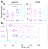
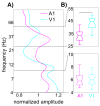
Attending to a stimulus enhances its neuronal representation, even at the level of primary sensory cortex. Cross-modal modulation can similarly enhance a neuronal representation, and this process can also operate at the primary cortical level. Phase reset of ongoing neuronal oscillatory activity has been shown to be an important element of the underlying modulation of local cortical excitability in both cases. We investigated the influence of attention on oscillatory phase reset in primary auditory and visual cortices of macaques performing an intermodal selective attention task. In addition to responses "driven" by preferred modality stimuli, we noted that both preferred and nonpreferred modality stimuli could "modulate" local cortical excitability by phase reset of ongoing oscillatory activity, and that this effect was linked to their being attended. These findings outline a supramodal mechanism by which attention can control neurophysiological context, thus determining the representation of specific sensory content in primary sensory cortex.
Figures


Comment in
-
Phase resetting as a mechanism for supramodal attentional control.Neuron. 2009 Nov 12;64(3):300-2. doi: 10.1016/j.neuron.2009.10.022. Neuron. 2009. PMID: 19914178
Similar articles
-
Phase resetting as a mechanism for supramodal attentional control.Neuron. 2009 Nov 12;64(3):300-2. doi: 10.1016/j.neuron.2009.10.022. Neuron. 2009. PMID: 19914178
-
The spectrotemporal filter mechanism of auditory selective attention.Neuron. 2013 Feb 20;77(4):750-61. doi: 10.1016/j.neuron.2012.11.034. Neuron. 2013. PMID: 23439126 Free PMC article.
-
Coupling between Theta Oscillations and Cognitive Control Network during Cross-Modal Visual and Auditory Attention: Supramodal vs Modality-Specific Mechanisms.PLoS One. 2016 Jul 8;11(7):e0158465. doi: 10.1371/journal.pone.0158465. eCollection 2016. PLoS One. 2016. PMID: 27391013 Free PMC article. Clinical Trial.
-
Look now and hear what's coming: on the functional role of cross-modal phase reset.Hear Res. 2014 Jan;307:144-52. doi: 10.1016/j.heares.2013.07.002. Epub 2013 Jul 12. Hear Res. 2014. PMID: 23856236 Review.
-
Defining Auditory-Visual Objects: Behavioral Tests and Physiological Mechanisms.Trends Neurosci. 2016 Feb;39(2):74-85. doi: 10.1016/j.tins.2015.12.007. Epub 2016 Jan 15. Trends Neurosci. 2016. PMID: 26775728 Free PMC article. Review.
Cited by
-
Looming signals reveal synergistic principles of multisensory integration.J Neurosci. 2012 Jan 25;32(4):1171-82. doi: 10.1523/JNEUROSCI.5517-11.2012. J Neurosci. 2012. PMID: 22279203 Free PMC article.
-
Predictive motor control of sensory dynamics in auditory active sensing.Curr Opin Neurobiol. 2015 Apr;31:230-8. doi: 10.1016/j.conb.2014.12.005. Epub 2015 Jan 13. Curr Opin Neurobiol. 2015. PMID: 25594376 Free PMC article. Review.
-
A precluding but not ensuring role of entrained low-frequency oscillations for auditory perception.J Neurosci. 2012 Aug 29;32(35):12268-76. doi: 10.1523/JNEUROSCI.1877-12.2012. J Neurosci. 2012. PMID: 22933808 Free PMC article. Clinical Trial.
-
Modulation of the Visual to Auditory Human Inhibitory Brain Network: An EEG Dipole Source Localization Study.Brain Sci. 2019 Aug 27;9(9):216. doi: 10.3390/brainsci9090216. Brain Sci. 2019. PMID: 31461954 Free PMC article.
-
Altered neural oscillations underlying visuospatial processing in cerebral visual impairment.Brain Commun. 2023 Aug 28;5(5):fcad232. doi: 10.1093/braincomms/fcad232. eCollection 2023. Brain Commun. 2023. PMID: 37693815 Free PMC article.
References
-
- Allman BL, Meredith MA. Multisensory processing in “unimodal” neurons: cross-modal subthreshold auditory effects in cat extrastriate visual cortex. J Neurophysiol. 2007;98:545–549. - PubMed
-
- Alsius A, Navarra J, Campbell R, Soto-Faraco S. Audiovisual integration of speech falters under high attention demands. Curr Biol. 2005;15:839–843. - PubMed
-
- Alsius A, Navarra J, Soto-Faraco S. Attention to touch weakens audiovisual speech integration. Exp Brain Res. 2007;183:399–404. - PubMed
-
- Basar E. EEG-brain dynamics: relation between EEG and Brain evoked potentials. Amsterdam: Elsevier; 1980.
Publication types
MeSH terms
Grants and funding
LinkOut - more resources
Full Text Sources
Other Literature Sources

