Review
. 2010 Feb;1804(2):245-62.
doi: 10.1016/j.bbapap.2009.11.004. Epub 2009 Nov 13.

The structural biochemistry of the superoxide dismutases

Affiliations
Review

The structural biochemistry of the superoxide dismutases

J J P Perry et al. Biochim Biophys Acta. 2010 Feb.

Abstract

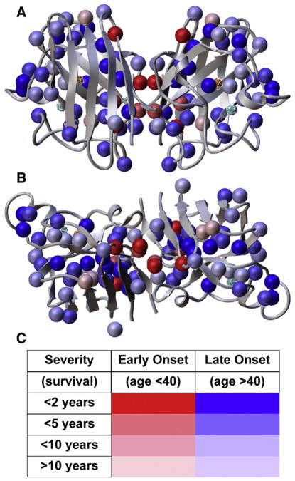
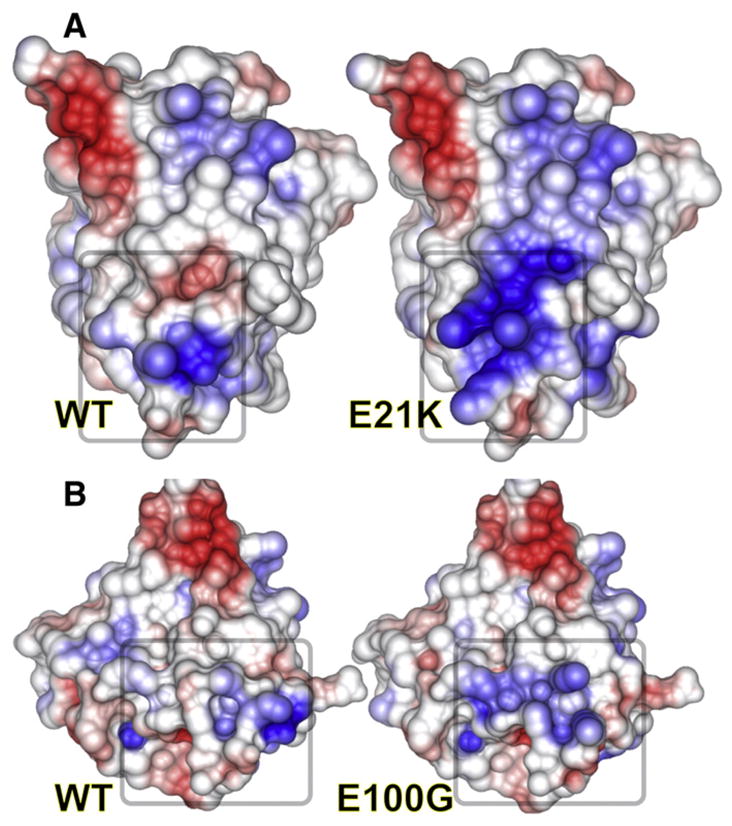
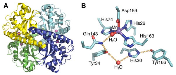
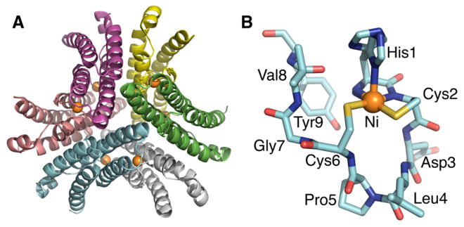
The discovery of superoxide dismutases (SODs), which convert superoxide radicals to molecular oxygen and hydrogen peroxide, has been termed the most important discovery of modern biology never to win a Nobel Prize. Here, we review the reasons this discovery has been underappreciated, as well as discuss the robust results supporting its premier biological importance and utility for current research. We highlight our understanding of SOD function gained through structural biology analyses, which reveal important hydrogen-bonding schemes and metal-binding motifs. These structural features create remarkable enzymes that promote catalysis at faster than diffusion-limited rates by using electrostatic guidance. These architectures additionally alter the redox potential of the active site metal center to a range suitable for the superoxide disproportionation reaction and protect against inhibition of catalysis by molecules such as phosphate. SOD structures may also control their enzymatic activity through product inhibition; manipulation of these product inhibition levels has the potential to generate therapeutic forms of SOD. Markedly, structural destabilization of the SOD architecture can lead to disease, as mutations in Cu,ZnSOD may result in familial amyotrophic lateral sclerosis, a relatively common, rapidly progressing and fatal neurodegenerative disorder. We describe our current understanding of how these Cu,ZnSOD mutations may lead to aggregation/fibril formation, as a detailed understanding of these mechanisms provides new avenues for the development of therapeutics against this so far untreatable neurodegenerative pathology.


Figures

Fig. 1
Fig. 1
Cu,ZnSOD sequence conservation, fold, and structural and functional regions. (A) Structure-based alignment of eukaryotic Cu,ZnSOD proteins with solved structures: HsSOD, H. sapiens; BtSOD, B. taurus; ApSOD, A. pompejana; XlSOD, X. laevis; SoSOD, S. oleracea; PaSOD, P. atrosanguina; SmSOD, S. mansoni; Sc, S. cerevisiae. Structural elements and secondary structure of HsSOD are noted above the alignment. Asterisks mark ALS sites in HsSOD. Letters below alignment: C, copper-binding ligand; D, disulfide cysteine; B, bridging histidine; Z, zinc-binding ligand; H, H2O2 liganding residue in ApSOD. (B) HsSOD structure 1PU0. Key structural elements in (A) are color coded with abbreviations (V-loop, Variable loop; GK1 and GK2, Greek key loops 1 and 2; S-S, Disulfide region). N and C denote termini. Metal-liganding residues are shown as sticks with the bridging histidine His61 in white.
Fig. 2
Fig. 2
(A) Electrostatic potential mapped onto the Alvinella pompejana surface from the ApSOD-H2O2 complex crystal structure. Electrostatic potential (blue positive, red negative) shows active site electrostatic attraction for superoxide anion. The H2O2 molecule (red spheres) occupies the position of the substrate/intermediate/product of the reaction. The spheres representing reduced copper (left) and zinc (right) are brought forward to show their relative positions. (B) Sliced version of the surface. From this view, the steric restrictions on the substrate/intermediate/product are readily recognized. Within the crystals, the copper ions (gold) were found in both the oxidized and reduced forms and show the short distance required for copper ion movement during SOD catalysis.
Fig. 3
Fig. 3
Distribution of ALS mutations within the HsSOD Structure. (A) SOD residues known to be mutated in ALS patients, shown with colored side chains and spheres for glycine Cα atoms. One subunit is yellow, the other orange. The majority of the β-barrel core and edge residues, and the dimer interface residues are ALS SOD mutation sites. These residues are important for maintaining β-barrel stability and quaternary structure. Many outer surface residues that are mutated in ALS patients contain side chain oxygen atoms potentially important for maintaining surface electrostatic potential or for stabilizing hydrogen bonding and salt-bridge interactions. (B) SOD residues not implicated in ALS, colored and shown from the same view as in (A). The active site (lower left) and the outer surface contain many residues not linked to ALS.
Fig. 4
Fig. 4
Disease onset and severity mapped onto ALS mutation sites in HsSOD structure. (A) FALS mutation sites (spheres) are found throughout the SOD sequence at sites expected to reduce the integrity of the β-barrel, especially the packing within the β-barrel core, “cork” regions, the dimer interface (center) and other framework features. (B) 90° rotation from (A) around horizontal axis. (C) Color codes for the spheres in (A) and (B) show the severity and age of onset linked to each mutation. Mutations linked to the most rapid and severe mutations are located within the dimer interface or in the β-barrel at positions likely to destabilize the dimer interface. The active site Cu and Zn ions are also shown as spheres within cages. Data from Wang et al. [63] were used to generate the figure.
Fig. 5
Fig. 5
Effects of single-point mutations in HsSOD on electrostatic potential. Electrostatic potential was mapped onto the surfaces of wild-type (left) vs. mutant (right) HsSOD models for the E21K and E100G HsSOD mutants. Loss of charge complementarity can dramatically change the electrostatic potential and reduce stability, both of which may result in aberrant SOD interactions.
Fig. 6
Fig. 6
Human MnSOD crystal structure. (A) The wild-type homotetrameric MnSOD structure (1LUV.pdb), containing two symmetrical four-helix bundles and four C-terminal α/β domains, is depicted with the four separate polypeptide chains colored in cyan, blue, green and yellow, and active site manganese ions depicted as magenta spheres. (B) MnSOD active site and hydrogen-bonding scheme. The side chains of active site residues His26, His74, His163, and Asp159 bind the Mn ion, in conjunction with a solvent molecule. A hydrogen-bonding network, depicted in orange spheres, is observed in the MnSOD active site, extending from the metal-bound solvent. The solvent forms a hydrogen bond to Gln143, and the network is continued with a hydrogen bond to Tyr34. A conserved water molecule then mediates the hydrogen bond between Tyr34 and His30, and His30 also forms a hydrogen bond with Tyr166 from an adjacent subunit.
Fig. 7
Fig. 7
S. coelicolor NiSOD crystal structure. (A) NiSOD forms a hexameric assembly (1T6U.pdb) of right-handed four-helix bundles that create a ~60 Å diameter structure with a ~20 Å deep central cavity. Extending from each four-helix bundle are N-terminal Ni-hooks, with the Ni ions displayed as orange spheres. (B) The nine-residue NiSOD metal-binding hook contains the conserved His-Cys-X-X-Pro-Cys-Gly-X-Tyr motif, which chelates a single metal ion and provides key residues for catalysis.

Similar articles

Cited by

References

    1. McCord JM, Fridovich I. Superoxide dismutase. An enzymic function for erythrocuprein (hemocuprein) J Biol Chem. 1969;244:6049–6055. - PubMed
    1. Fridovich I. Superoxide anion radical (O2-.), superoxide dismutases, and related matters. J Biol Chem. 1997;272:18515–18517. - PubMed
    1. Lane N. Oxygen: The Molecule that Made the World. Oxford University Press; USA: 2003.
    1. Fridovich I. Superoxide dismutases. Adv Enzymol Relat Areas Mol Biol. 1986;58:61–97. - PubMed
    1. Babior BM, Kipnes RS, Curnutte JT. Biological defense mechanisms. The production by leukocytes of superoxide, a potential bactericidal agent. J Clin Invest. 1973;52:741–744. - PMC - PubMed

Publication types

Substances

LinkOut - more resources