. 2009 Oct;18 Suppl 3(Suppl 3):S10-7.
doi: 10.1016/S0960-9776(09)70266-8.

New hypotheses and opportunities in endocrine therapy: amplification of oestrogen-induced apoptosis

Affiliations

New hypotheses and opportunities in endocrine therapy: amplification of oestrogen-induced apoptosis

V Craig Jordan et al. Breast. 2009 Oct.

Abstract

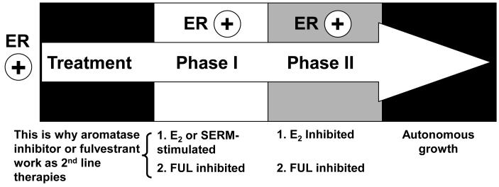
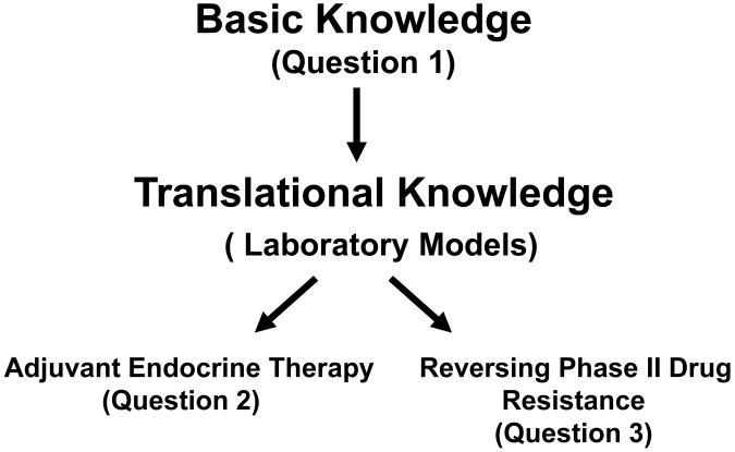
Aims: To outline the progress being made in the understanding of acquired resistance to long term therapy with the selective oestrogen receptor modulators (SERMs, tamoxifen and raloxifene) and aromatase inhibitors. The question to be addressed is how we can amplify the new biology of oestrogen-induced apoptosis to create more complete responses in exhaustively antihormone treated metastatic breast cancer.

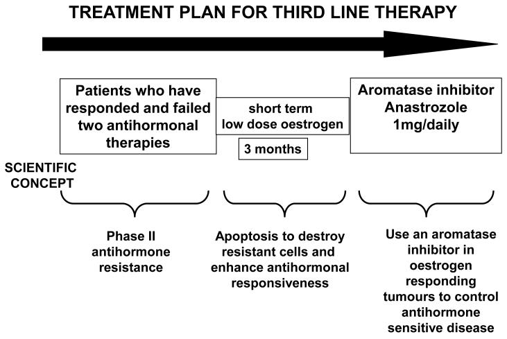
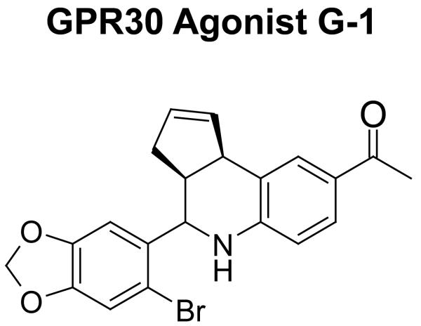
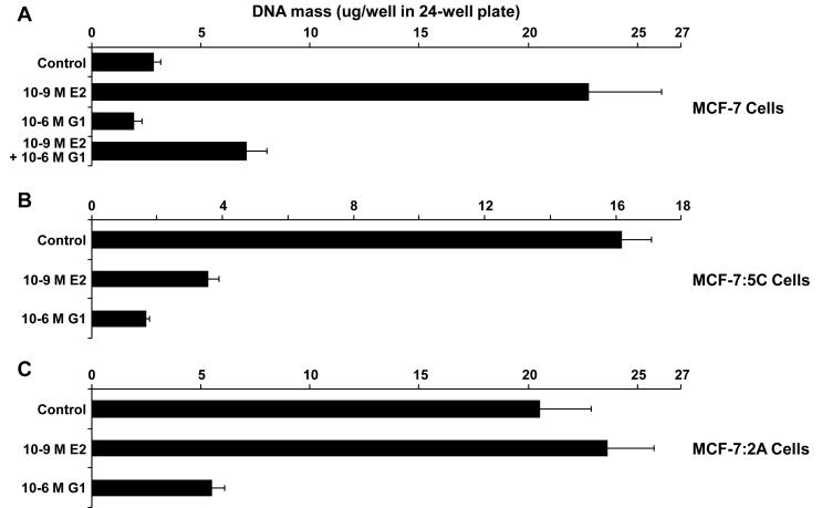
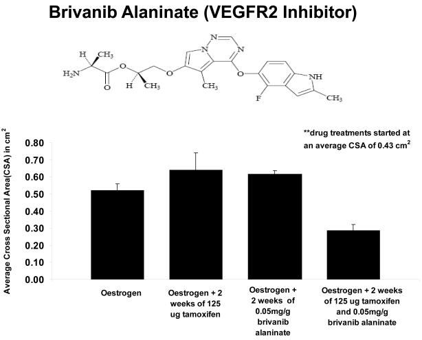
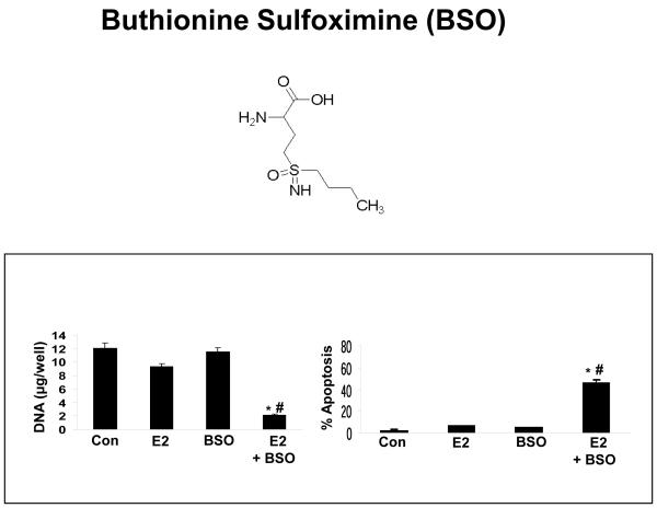
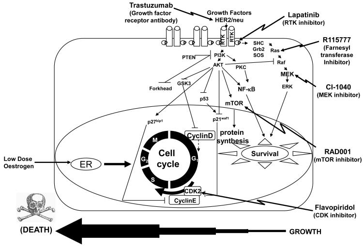
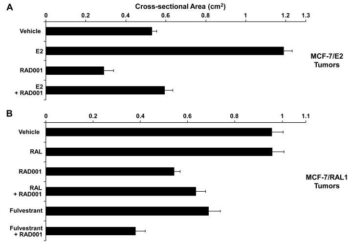
Methods and results: Three questions are posed and addressed. (1) Do we know how oestrogen works? (2) Can we improve adjuvant antihormonal therapy? (3) Can we enhance oestrogen-induced apoptosis? The new player in oestrogen action is GPR30 and there are new drugs specific for this target to trigger apoptosis. Similarly, anti-angiogenic drugs can be integrated into adjuvant antihormone therapy or to enhance oestrogen-induced apoptosis in Phase II antihormone resistant breast cancer. The goal is to reduce the development of acquired antihormone resistance or undermine the resistance of breast cancer cells to undergo apoptosis with oestrogen respectively. Finally, drugs to reduce the synthesis of glutathione, a subcellular molecule compound associated with drug resistance, can enhance oestradiol-induced apoptosis.

Conclusions: We propose an integrated approach for the rapid testing of agents to blunt survival pathways and amplify oestrogen-induced apoptosis and tumour regression in Phase II resistant metastatic breast cancer. This Pharma platform will provide rapid clinical results to predict efficacy in large scale clinical trials.


Figures

Figure 1
Figure 1
The evolution of drug resistance to SERMs. Acquired resistance occurs during long-term treatment with a SERM and is evidenced by SERM-stimulated breast tumour growth. Tumours also continue to exploit oestrogen for growth when the SERM is stopped, so a dual signal transduction process develops. The aromatase inhibitors prevent tumour growth in SERM-resistant disease and fulvestrant that destroys the ER is also effective. This phase of drug resistance is referred to as Phase I resistance. Continued exposure to a SERM results in continued SERM-stimulated growth (Phase II), but eventually autonomous growth occurs that is unresponsive to fulvestrant or aromatase inhibitors. The event that distinguishes Phase I from Phase II acquired resistance is a remarkable switching mechanism that now causes apoptosis, rather than growth, with physiologic levels of oestrogen. These distinct phases of laboratory drug resistance (20, 25) have their clinical parallels and this new knowledge is being integrated into the treatment plan.
Figure 2
Figure 2
Clinical protocol to investigate the efficacy of oestradiol induced apoptosis in long-term endocrine refractory breast cancer. An anticipated treatment plan for third-line endocrine therapy. Patients must have responded and experience treatment failure with two successive antihormone therapies to be eligible for a course of low-dose oestradiol therapy for 3 months. The anticipated response rate is 30% and responding patients will be treated with anastrozole until relapse. Validation of the treatment plan will establish a platform to enhance response rates with apoptotic oestrogen by integrating known inhibitors of tumour survival pathways into the 3-month debulking “oestrogen purge”. The overall goal is to increase response rates and maintain patients for longer on antihormone strategies before chemotherapy is required.
Figure 3
Figure 3
The interactive translational research model employed to address new hypotheses and opportunities to amplify oestrogen-induced apoptosis for the treatment of Phase II endocrine resistant breast cancer. The questions posed are described in the text.
Figure 4
Figure 4
G1, the first of a new class of agents that act as selective agonists of GPR30. A range of antagonists is also being developed.
Figure 5
Figure 5
The selective GPR30 agonist G1 inhibits growth of (A) wild-type MCF-7 cells and of oestrogen deprivation-resistant (B) MCF-7:5C and (C) MCF-7:2A cells. Cells were cultured under oestrogen-free conditions for 4 days, and then seeded into 24-well plates. Wild-type MCF-7 cells were seeded at 15,000 cells per well, MCF-7:5C cells at 25,000 cells per well, and MCF-7:2A cells at 30,000 cells per well. Beginning 24 hours after seeding (day 0) and every 2 days thereafter up to 6 days (days 2, 4, and 6), the cells were treated with 1 nM E2, 1 μM G1, 1 nM E2 + 1 μM G1, or Control (0.1% EtOH)-treated. The experiment was stopped on day 7. As a measure of proliferation, the amount of DNA per well was determined using a fluorescence-based DNA quantitation assay (CyQuant GR, Invitrogen, Carlsbad, CA). Data are shown as the mean of 8 replicate wells per group ± SD. (A) In wild-type MCF-7 cells, G1 significantly inhibited E2-stimulated growth by 78% (E2 vs. E2+G1, P < 0.0001), and inhibited growth relative to control-treated cells (control vs. G1, P = 0.0003). (B) In estrogen deprivation-resistant MCF-7:5C cells, E2 induced apoptosis as expected leading to a 78% reduction in growth (control vs. E2, P < 0.0001). G1 also significantly inhibited growth by 90% (control vs. G1, P < 0.0001), and further, was more potent than E2 (G1 vs. E2 P < 0.0001). (C) The oestrogen deprivation-resistant MCF-7:2A cells grew independently of E2 within the 7 day course of the experiment, as expected, yet G1 significantly inhibited growth by 73% (P < 0.0001).
Figure 6
Figure 6
Established MCF-7 E2 tumours and their response to various drug treatments. Tumours were implanted bilaterally into the mammary fat pads of athymic mice and 0.3 cm estradiol capsules were implanted subcutaneously into the dorsum of each mouse. Tumours were grown to 0.43 cm^2 and then drug treatments were initiated. Tumours that were treated with 125 ug of tamoxifen or 0.05 mg/g brivanib alaninate were unable to overcome oestradiol stimulated growth (p=0.65, p=0.21). . Tumours continued to grow in the presence of oestrogen. When 125 ug of tamoxifen was combined with 0.05 mg/g brivanib alaninate, the effect was synergistic (p=.009) and the tumours decreased in size. The tumours were 38% smaller than the oestrogen treated tumours, even though the observed difference was not significant (p=0.16). However, the decrease in average cross sectional area was significant when comparing the combination treatment to tamoxifen treated tumours (p=.01) or those treated with brivanib alaninate (p=007).
Figure 7
Figure 7
The combination treatment of BSO plus oestradiol inhibits the growth of antihormone-resistant MCF-7:2A breast cancer cells. MCF-7:2A cells (30,000/well) were seeded in 24-well plates and after 24 hours were treated with < 0.1% ethanol vehicle (control), 1 nM E2 (E2), 100 μM BSO (chemical structure shown above), or 100 μM BSO plus 1 nM E2 for 7 days. At the indicated time point, cells were harvested and total DNA (μg/well) was quantitated as described in Materials and Methods. The data represent the mean of three independent experiments; bars, ±SE. **, P < 0.001 compared with control cells; ##, P < 0.001 compared with oestradiol-treated cells. Annexin V staining for apoptosis was performed in MCF-7:2A cells following BSO plus E2 treatment. Quantitation of apoptosis (percent of control) in the different treatment groups is shown on the right. bars, ±SEs. *, P < 0.05 compared with control cells; #, P < 0.01 compared with oestradiol-treated cells.
Figure 8
Figure 8
Hypothetical apoptosis enhancement strategy to amplify the tumouricidal action of low dose oestradiol treatment. The strategy is to employ targeted agents from the pharmacological industry to block several pathways and shift the cellular equilibrium to apoptosis in oestrogen refractory Phase II resistant cells. The diagram illustrates candidate drugs to create a cocktail in the proposed Pharma platform. Drugs would be tested singly with oestradiol against alone or in increasing combinations.
Figure 9
Figure 9. Growth inhibition of naïve MCF-7/ E2 tumours and SERM-resistant MCF-7/RAL1 tumours in response to RAD001 (everolimus)
(A) RAD001 inhibition of MCF-7/ E2 tumour growth. Twenty ovariectomized athymic nude mice were bilaterally transplanted with MCF-7/ E2 tumour pieces 1 mm3 in size in the axillary mammary fat pads, and implanted with a 0.3 cm E2 silastic capsule sc. Once the tumours grew to an average cross-sectional area of 0.39 cm2, the animals were randomized into 4 treatment groups of 5 mice per group (10 tumours per group) corresponding to Vehicle (of the RAD001 formulation), E2 (0.3 cm E2 capsule sc), RAD001 [40 mg/kg/day (6.25 mg/day) RAD001 given 5 days/week], and E2 + RAD001 (0.3 cm E2 capsule sc plus 6.25 mg/day RAD001 given 5 days/week). The average cross-sectional area of RAD001-treated MCF-7/ E2 tumours was significantly smaller than Vehicle-treated tumours (P = 0.0066, T test). Similarly, the average cross-sectional are a of E2 + RAD001-treated tumours was significantly smaller than E2 alone -treated tumours (P < 0.0001). (B) RAD001 inhibition of MCF-7/RAL1 tumour growth. Thirty ovariectomized athymic nude mice were bilaterally implanted in the axillary mammary fat pads with 1 mm3 MCF-7/RAL1 tumour pieces. Mice were treated with 1.5 mg/day RAL po until the MCF-7/RAL1 tumours grew to an average cross-sectional area of 0.26 cm2, and then the animals were separated into 6 treatment groups of 5 mice each (10 tumours per group) corresponding to Vehicle (of the RAD001 formulation), 1.5 mg/day RAL po, RAD001 (6.25 mg/day RAD001 given 5 days/week), RAL + RAD001 (1.5 mg/day RAL po plus 6.25 mg/day RAD001 given 5 days/week), Fulvestrant (2 mg/day sc of the clinically used Faslodex preparation given 5 days/week), Fulvestrant + RAD001 (2 mg/day Faslodex sc plus 6.25 mg/day RAD001 given 5 days/week). The average cross-sectional areas of RAD001-treated and Fulvestrant-treated tumours were each significantly smaller than Vehicle-treated tumours (P < 0.0001 and P = 0.0015, respectively). Similarly, RAL + RAD001 –treated tumours were significantly smaller than RAL-treated tumours (P = 0.0002). Additionally, Fulvestrant + RAD001 –treated tumors were significantly smaller than RAD001 alone –treated tumors (P = 0.0026) or Fulvestrant alone –treated tumors (P = 0.0004). The data shown represent the average cross-sectional tumour area (cm2) per group ± SE. Tumor cross-sectional area was calculated using the equation: (l/2) × (w/2) × π. The cross-sectional areas of MCF-7/E2 tumours were compared at day 41, and of MCF-7/RAL1 tumours at day 54.

Similar articles

Cited by

References

    1. Jordan VC. Tamoxifen: a most unlikely pioneering medicine. Nature Reviews Drug Discovery. 2003;2:205–13. - PubMed
    1. Cole MP, Jones CT, Todd ID. A new anti-oestrogenic agent in late breast cancer. An early clinical appraisal of ICI 46474. Br J Cancer. 1971;25:270–5. - PMC - PubMed
    1. Ward HW. Anti-oestrogen therapy for breast cancer: a trial of tamoxifen at two dose levels. Br Med J. 1973;1:13–4. - PMC - PubMed
    1. Ingle JN, Ahmann DL, Green SJ, Edmonson JH, Bisel HF, Kvols LK, et al. Randomized clinical trial of diethylstilbestrol versus tamoxifen in postmenopausal women with advanced breast cancer. N Engl J Med. 1981;304:16–21. - PubMed
    1. Jordan VC. Tamoxifen: catalyst for the change to targeted therapy. Eur J Cancer. 2008;44:30–8. - PMC - PubMed

Publication types

MeSH terms