. 2010;261(1):9-22.
doi: 10.1016/j.cellimm.2009.10.004. Epub 2009 Oct 19.

BAFF overexpression promotes anti-dsDNA B-cell maturation and antibody secretion

Affiliations

BAFF overexpression promotes anti-dsDNA B-cell maturation and antibody secretion

Mitchell Thorn et al. Cell Immunol. 2010.

Abstract

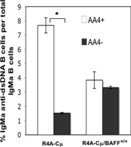
Overexpression of BAFF is believed to play an important role in systemic lupus erythematosus and elevated levels of serum BAFF have been found in lupus patients. Excess BAFF also leads to overproduction of anti-dsDNA antibodies and a lupus-like syndrome in mice. In the present study, we use mice transgenic for the R4A-Cmu (IgM) heavy chain of an anti-dsDNA antibody, to study the effects of BAFF overexpression on anti-dsDNA B-cell regulation. We observe that overexpression of BAFF promotes anti-dsDNA B-cell maturation and secretion of antibody and enriches for transgenic anti-dsDNA B cells in the marginal zone and follicular splenic compartments. In addition, our data suggests that BAFF rescues a subset of anti-dsDNA B cells from a regulatory checkpoint in the transitional stage of development.


Conflict of interest statement

There are no commercial or financial conflicts of interest associated with this study.

Figures

Figure 1
Figure 1. R4A-Cμ mice homozygous for BAFF secrete elevated levels of IgMa anti-dsDNA antibody
A. Sera from 13 R4A-Cμ mice, 18 R4A-Cμ/BAFF+/- mice and 18 R4Cμ/BAFF+/+ mice were tested by ELISA for Tg, IgMa anti-dsDNA antibody. Horizontal lines represent average OD at 405 nm. Dotted line represents 4 standard deviation units above the average OD of serum samples from R4A-Cμ mice. B. IgMa anti-dsDNA antibody concentrations were measured from the sera of 10 R4A-Cμ mice (circles), R4A-Cμ/BAFF+/- mice (squares) and R4Cμ/BAFF+/+ mice (triangles) by a quantitative ELISA.
Figure 2
Figure 2. R4A-Cμ anti-dsDNA B cells require high concentration of serum BAFF for the secretion of antibody
A. Variations in serum BAFF concentrations in mice overexpressing BAFF. Serum concentrations of BAFF were measured by ELISA for 11 R4A-Cμ mice, 15 R4A-Cμ/BAFF+/- and 15 R4A-Cμ/BAFF+/+ mice. Horizontal bars represent average concentrations of BAFF. B. A higher concentration of serum BAFF is required for the secretion of Tg versus non Tg anti-dsDNA antibody. Serum BAFF concentrations were measured for 30 R4A-Cμ/BAFF+/+ mice. Mice were grouped into those with high serum concentrations of BAFF (300 - 1000 ng/ml) and those with lower concentrations of BAFF (10 - 250 ng/ml). Graph depicts percent of mice in each group secreting either non Tg (IgMb or IgG) (open bar) or Tg (IgMa) anti-dsDNA antibody (closed bar).
Figure 3
Figure 3. Expression levels of BR3 and TACI are reduced on R4A-Cμ, Tg B cells
Splenocytes from R4A-Cμ/BAFF+/+ mice were immunostained with antibodies to IgMa, IgMb, B220, AA4.1, CD23, TACI, and BAFF-R. Expression levels of BR3 (A) or TACI (B) were examined on transitional (B22O+ AA4+), FO (AA4-, CD23hi IgMint), and MZ (AA4- CD23lo IgMhi) B cells expressing the IgMa transgene or endogenous IgMb. Gates were set on IgMa (solid black line) or IgMb (solid grey line) B cells with a transitional, FO, or MZ phenotype. Dashed histogram indicates isotype control. Dotted vertical line depicts arbitrary reference point. Histograms are representative of at least 6 experiments. C. Ratios of the Mean Fluorescent Intensities (MFIs) of BR3 (in A) to TACI (in B).
Figure 4
Figure 4. BAFF overexpression does not alter B cell development in the bone marrow
A. Bone marrow B cells from R4A-Cμ, R4A-Cμ/BAFF+/+, WT, and BAFF mice were immunostained with antibodies to B220, IgM and CD43 and analyzed by flow cytometry. Gates were set on B220+ cells. Insert indicates windows for pro (CD43+IgM-), pre (CD43-IgM-), and immature/mature IgM (CD43- IgM+) B cells. B. Splenic B cells from R4A-Cμ, R4A-Cμ/BAFF+/+, WT, and BAFF mice were immunostained with antibodies to B220 and IgM. Gates were set on live lymphocytes. C. Splenic B cells from R4A-Cμ and R4A-Cμ/BAFF+/+ mice were immunostained with antibodies to B220 and IgMa. Results in A, B, and C are representative of at least 3 experiments.
Figure 5
Figure 5. Overexpression of BAFF promotes the maturation of R4A-Cμ, Tg B cells in the periphery
Splenocytes from R4A-Cμ and R4A-Cμ/BAFF+/+ mice were immunostained with antibodies to IgMa, B220, CD24, and AA4.1 and analyzed by flow cytometry. A. Histogram displays CD24hi and CD24lo IgMa B cells. Gate set on live, IgMa, B cells. B. Contour graph indicates frequency of AA4.1+ and AA4.1- Tg B cells in R4A-Cμ and R4A-Cμ/BAFF mice. Gate set on live, IgMa, B cells. C. Splenocytes from R4A-Cμ mice were sorted to collect AA4.1+ Tg B cells, using a BD FACS, ARIA. Purity of the post sort population was 95% (right panel). D. Sorted AA4.1+ IgMa cells from C were transferred into wild type or BAFF mice, intravenously. Two days later, the spleens of recipient mice were harvested and immunostained with antibodies to IgMa and AA4.1 to assess the maturation state of the transferred cells. Results in A and B are representative of 6 experiments and results in C and D are representative of 3 experiments.
Figure 6
Figure 6. Increased frequencies of late transitional and MZ, Tg B cells in R4A-Cμ/BAFF+/+ mice
Splenocytes from R4A-Cμ and R4A-Cμ/BAFF+/+ mice were immunostained with antibodies to IgMa, B220, CD23, and AA4.1 and analyzed by flow cytometry. A. Contour graph indicates the frequency of AA4.1+, T1 (IgMa hi, CD23-), T2 (IgMa hi, CD23+) and T3 (IgMa lo, CD23+) B cells in R4A-Cμ and R4A-Cμ/BAFF+/+ mice. Gates were set on live, IgMa AA4.1+ B cells. B. Splenocytes from R4A-Cμ and R4A-Cμ/BAFF+/+ mice (top panel), and WT and BAFF mice (bottom panel) were immunostained with antibodies to B220, CD21, CD23, and antibody to IgMa (to detect Tg B cells, top panel) or antibody to IgMb (to detect non Tg B cells from WT and BAFF mice, bottom panel) and analyzed by flow cytometry to determine the frequency of MZ (CD21hi CD23lo) and FO (CD21int CD23hi) B cells. Gates were set on live B220 IgMa cells for R4A-Cμ and R4A-Cμ/BAFF+/+ mice and live B220 IgMb cells for WT and BAFF Tg mice. Results are representative of 6 experiments.
Figure 7
Figure 7. MZ and FO compartments are enriched with Tg IgMa B cells in R4A-Cμ/BAFF+/+ mice
Frozen spleen sections (6 μm) from R4A-Cμ and R4A-Cμ/BAFF+/+ mice were immunostained with antibody to MOMA-1 (marker for metallophilic macrophages) and antibodies to total IgM (A) or IgMa (B). Immunostaining of marginal sinus is indicated by green fluorescence. Immunostaining of total IgM B cells in (A) or IgMa B cells in (B) is depicted by red fluorescence. (PALS, Periarteriolar lymphatic sheath; MZ, marginal zone: FO, follicle). Crossbars indicate width of the MZ. Sections are representative of 4 experiments.
Figure 8
Figure 8. The number of R4A-Cμ, Tg anti-dsDNA B cells is increased in the MZ and FO of R4A-Cμ/BAFF+/+ mice
Splenic B cells from R4A-Cμ and R4A-Cμ/BAFF+/+ mice were immunostained with antibodies to B220, CD21, and CD23 and sorted to obtain MZ (B220+ CD21hi CD23lo) and FO (B220+ CD21int CD23hi)B cells. Cells were then cultured in either media alone or activated in vitro with 1μg/ml of stimulatory CpG oligonucleotides for 48 hours and then IgMa anti-dsDNA secreting B cells (A) and total IgMa secreting B cells (B) were enumerated by ELIspot.
Figure 9
Figure 9. BAFF enables R4A-Cμ, Tg anti-dsDNA B cells to escape the transitional stage of development
Splenic B cells from R4A-Cμ and R4A-Cμ/BAFF+/+ mice were immunostained with antibodies to B220 and AA4.1 and sorted into B220+ AA4+ (transitional) and B220+ AA4- (mature) populations. They were then activated in vitro with 1 μg/ml of stimulatory CpG oligonucleotides and the number of IgMa anti-dsDNA secreting B cells and the total number of IgMa secreting B cells was determined by ELIspot. Results are represented as the percent of IgMa secreting B cells that are specific for dsDNA. * indicates p< 0.005.
Figure 10
Figure 10. Negligible frequency of peritoneal B1 cells express the IgMa transgene in R4A-Cμ/BAFF+/+ mice
Peritoneal cells isolated by lavage from R4A-Cμ and R4A-Cμ/BAFF+/+ mice were immunostained with antibodies to B220, CD5, and IgMa to detect the frequency of B1 cells expressing the transgene and IgMb to detect B1 cells expressing non Tg IgM and examined by flow cytometry. A. The frequency of B220+ CD5+ B cells is >3 fold greater in R4A-Cμ/BAFF+/+ than R4-Cμ mice. B. Very few peritoneal B cells derived from the B1 lineage express the IgMa transgene in both R4A-Cμ and R4A-Cμ/BAFF+/+ mice (top panel). Most express the non Tg IgMb (bottom panel). Gate was set on B220+ B cells and the frequencies of CD5+ and CD5- B cells expressing IgMa (top panels) or IgMb (lower panels) are indicated.

Similar articles

Cited by

References

    1. Batten M, Groom J, Cachero TG, Qian F, Schneider P, Tschopp J, Browning JL, Mackay F. BAFF mediates survival of peripheral immature B lymphocytes. J Exp Med. 2000;192:1453–1465. - PMC - PubMed
    1. Rolink AG, Tschopp J, Schneider P, Melchers F. BAFF is a survival and maturation factor for mouse B cells. Eur J Immunol. 2002;32:2004–2010. - PubMed
    1. Schiemann B, Gommerman JL, Vora K, Cachero TG, Shulga-Morskaya S, Dobles M, Frew e, Scott ML. An essential role for BAFF in the normal development of B cells through a BCMA-independent pathway. Science. 2001;293:2111–2114. - PubMed
    1. Schneider P, Takatsuka H, Wilson A, Mackay F, Tardivel A, Lens S, Cachero TG, Finke D, Beermann F, Tschopp J. Maturation of Marginal zone and Follicular B cells requires B cell activating factor of the tumor necrosis factor family and is independent of B cell maturation antigen. J Exp Med. 2001;194:1691–1697. - PMC - PubMed
    1. Gorelik L, Gilbride K, Dobles M, Kalled SL, Zandman D, Scott ML. Normal B cell homeostasis requires B cell activation factor production by radiation-resistant cells. J Exp Med. 2003;198:937–945. - PMC - PubMed

Publication types

LinkOut - more resources