

Endodermal origin of bladder trigone inferred from mesenchymal-epithelial interaction
- PMID: 19914648
- PMCID: PMC2794964
- DOI: 10.1016/j.juro.2009.08.107
Endodermal origin of bladder trigone inferred from mesenchymal-epithelial interaction
Abstract
Purpose: In the classic view of bladder development the trigone originates from the mesoderm derived wolffian ducts while the remainder of the bladder originates from the endoderm derived urogenital sinus. Recent molecular developmental studies have questioned the veracity of this received wisdom, suggesting an endodermal origin for the trigone. To shed further light on this issue we observed mesenchymal-epithelial interactions between trigone epithelium and fetal urogenital sinus mesenchyma to infer the trigonal germ layer of origin.
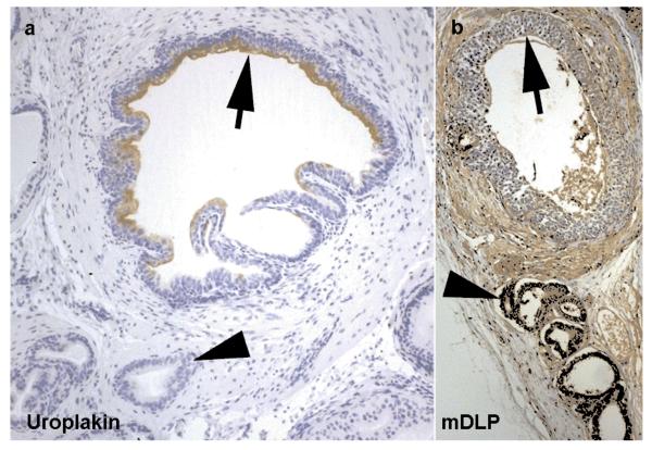
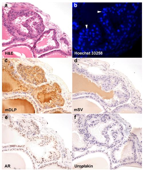
Materials and methods: Mouse trigone epithelium was recombined with fetal rat urogenital sinus mesenchyma in tissue recombinant grafts that were placed beneath the renal capsule of athymic mouse hosts. Grafts were harvested at 4 weeks. Control grafts with bladder dome and ureteral epithelium were also examined. Tissues were evaluated with hematoxylin and eosin, and Hoechst dye 33258 to confirm cell species origin. Immunohistochemistry was done with androgen receptor, broad spectrum uroplakin, dorsolateral prostate secretions and seminal vesicle secretions to differentiate prostatic and seminal vesicle differentiation.
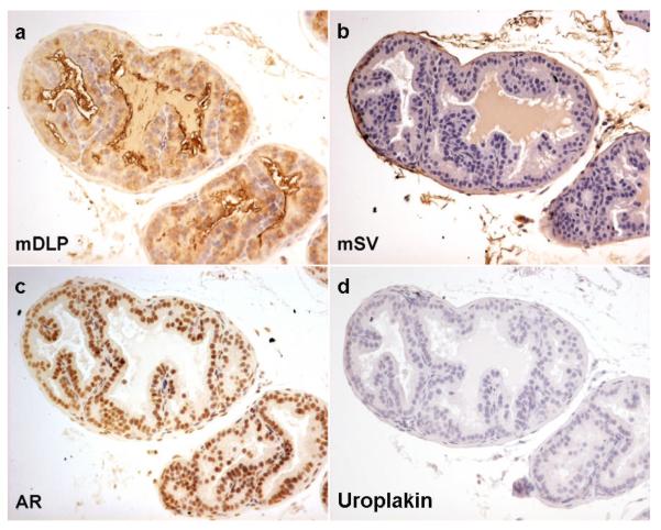
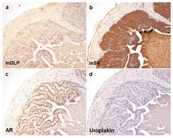
Results: Grafts of mouse trigone epithelium with fetal rat urogenital sinus mesenchyma yielded epithelial tissue that stained for dorsolateral prostate secretions but not for seminal vesicle secretions. Control grafts of bladder dome epithelium yielded the expected endodermal prostate differentiation. Control grafts of ureteral epithelium yielded the expected mesodermal seminal vesicle differentiation.
Conclusions: The consistent finding of prostatic epithelium in tissue recombinants of trigone epithelium and fetal urogenital sinus mesenchyma reinforces the hypothesis that the trigone is derived from the endoderm and not from the mesoderm, as commonly accepted.
Figures
References
-
- Wesson MB. Anatomical, embryological and physiological studies of the trigone and neck of the bladder. Journal of Urology. 1920;4:279.
-
- Batourina E, Tsai S, Lambert S, Sprenkle P, Viana R, Dutta S, et al. Apoptosis induced by vitamin A signaling is crucial for connecting the ureters to the bladder. Nat Genet. 2005;37:1082. - PubMed
-
- Viana R, Batourina E, Huang H, Dressler GR, Kobayashi A, Behringer RR, et al. The development of the bladder trigone, the center of the anti-reflux mechanism. Development. 2007;134:3763. - PubMed
-
- Cunha GR, Ricke W, Thomson A, Marker PC, Risbridger G, Hayward SW, et al. Hormonal, cellular, and molecular regulation of normal and neoplastic prostatic development. J Steroid Biochem Mol Biol. 2004;92:221. - PubMed
-
- Kurzrock EA, Baskin LS, Cunha GR. Ontogeny of the male urethra: theory of endodermal differentiation. Differentiation. 1999;64:115. - PubMed
