Review
. 2009 Dec;19(6):650-7.
doi: 10.1016/j.sbi.2009.10.017. Epub 2009 Nov 14.

Signaling mechanisms of the Mycobacterium tuberculosis receptor Ser/Thr protein kinases

Affiliations
Review

Signaling mechanisms of the Mycobacterium tuberculosis receptor Ser/Thr protein kinases

Tom Alber. Curr Opin Struct Biol. 2009 Dec.

Abstract

Like eukaryotes, bacteria express receptor Ser/Thr protein kinases (STPKs) that initiate a wide variety of signaling networks. Recent biochemical and structural studies of the STPKs of Mycobacterium tuberculosis have revealed that bacterial and eukaryotic STPKs adopt common folds and share mechanisms of substrate recognition and regulation. Mycobacterial receptor STPKs are activated by dimerization through two distinct interfaces that promote activation-loop phosphorylation. The active STPKs phosphorylate diverse substrates within the bacterial cell, including other kinases as well as proteins involved in many central physiological processes. In the case of the FHA-domain protein, GarA, the unphosphorylated protein regulates primary metabolism, while phosphorylation mediates GarA autoinhibition. These studies have begun to define the activation mechanisms and the biological regulatory functions of the mycobacterial STPKs.


Figures

Figure 1
Figure 1. The PknB KD:ATPγS structure resembles the transition-state analog complex of cAMP-dependent protein kinase (PKA)
Superimposed backbone ribbons show the similarity of the classic kinase folds of PknB (blue) and PKA (yellow), including the two-domain structure and the P-loop covering the nucleotide. The bound nucleotides and several active-site residues in PknB:ATP-γ-S (blue) and the PKA:ADP:AlF3:peptide transition-state-analog complex [45] shows that conserved residues contact the nucleotides in similar conformations.
Figure 2
Figure 2. Mtb PknB, PknE and human PKR KDs form structurally similar activated dimmers
Ribbon drawings of PknB-ATPγS (left; C-helix yellow, nucleotide in spheres), apo-PknE (middle), and a superposition (right) of PknB (blue) and PKR (orange) reveal the conserved architecture. The N-lobe interaction holds the catalytic sites away from each other and activates the unphosphorylated kinases by an allosteric mechanism. The back-to-back interface communicates to the active site over 25 Å across the protein.
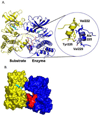
Fig. 3
Fig. 3. Inhibitor complex of a PknB KD mutant resembled an autophosphorylation complex
A. PknB monomers formed an asymmetric offset dimer with contacts between the G-helices. Identical interface residues occur in distinct environments in the presumptive substrate (yellow) and enzymatic (blue). The activation loop was disordered in the substrate KD (yellow dashed line) and ordered (red line) in the enzyme KD.) B. Surface representation showing the contacts between kinase molecules and the ordered activation loop (red) in the enzyme KD.
Fig. 4
Fig. 4. Model for Mtb receptor STPK regulation [24]
The inactive, unphosphorylated kinase monomer (left) is paired by an extracellular signal. The dimer adopts a conformation that is activated for autophosphorylation and substrate phosphorylation. The autophosphorylated monomers are active and amplify the signal (right). After the signal dissipates, the PstP phosphatase returns the kinase to the unphosphorylated inactive form.
Figure 5
Figure 5. Phosphorylation autoinhibits GarA
A. Schematic model of Mtb GarA inhibition [8]. The FHA domain (orange) engages the N-terminal tail (red) phosphorylated on Thr21 (by PknG) or Thr22 (by PknB), stabilizing a conformation that blocks binding of the STPKs and target enzymes GDH, KGD and GS. The N-terminal tail adopts many conformations in the on state. B. Ribbon diagram of the average NMR structure of GarA [8] showing the N-terminal tail (red) occluding the enzyme-binding surface. Spheres show pThr22 (atom colors) and sites where mutations block binding of all three (blue) or two of the three (light blue) unphosphorylated target enzymes. C. Superposition of the average NMR structures of OdhI [41], the GarA homolog in C. glutamicum, unphosphorylated (cyan) and phosphorylated on Thr15 (blue). Folding of the N-terminal tail into the pThr binding site causes little change in the core FHA domain structure.

References

    1. Cheek S, Ginalski K, Zhang H, Grishin NV. A comprehensive update of the sequence and structure classification of kinases. BMC Struct Biol. 2005;5:6. - PMC - PubMed
    1. Mougous JD, Gifford CA, Ramsdell TL, Mekalanos JJ. Threonine phosphorylation post-translationally regulates protein secretion in Pseudomonas aeruginosa. Nat Cell Biol. 2007;9:797–803. Genetic and biochemical analysis showed the central roles of the PpkA STPK, the antagonistic PppA Ser/Thr phosphatase and the Fha1 FHA-domain protein in assembling the type VI secretion apparatus in P. aeruginosa.

    1. Hsu F, Schwarz S, Mougous JD. TagR promotes PpkA-catalysed type VI secretion activation in Pseudomonas aeruginosa. Mol Microbiol. 2009;72:1111–1125. A fascinating study of type VI secretion in P. aeruginosa established a cascade of assembly reactions triggered by activation of the PpkA STPK by dimerization in vivo.

    1. S Shah IM, Laaberki MH, Popham DL, Dworkin J. A eukaryotic-like Ser/Thr kinase signals bacteria to exit dormancy in response to peptidoglycan fragments. Cell. 2008;135:486–496. A beautiful demonstration that peptidoglycan fragments signal through PknB orthologs, and the species specificity of signaling is established by the correspondence between the extracellular PASTA domains of the STPK and chemical variations in peptidoglycan.

    1. Av-Gay Y, Everett M. The eukaryotic-like Ser/Thr protein kinases of Mycobacterium tuberculosis. Trends Microbiol. 2000;8:238–244. The classic review of the Mtb kinome.

Publication types

MeSH terms

Substances

LinkOut - more resources