. 2010 Jan;9(1):97-106.
doi: 10.1128/EC.00203-09. Epub 2009 Nov 13.

RNA interference silencing of a major lipid droplet protein affects lipid droplet size in Chlamydomonas reinhardtii

Affiliations

RNA interference silencing of a major lipid droplet protein affects lipid droplet size in Chlamydomonas reinhardtii

Eric R Moellering et al. Eukaryot Cell. 2010 Jan.

Abstract

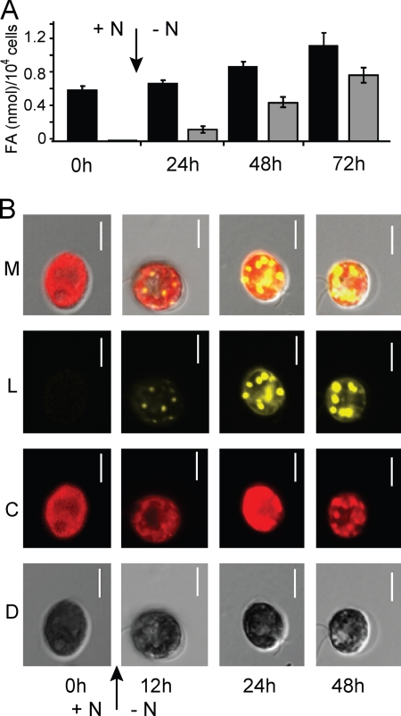
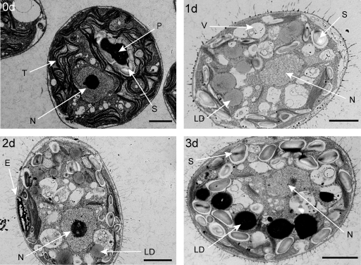
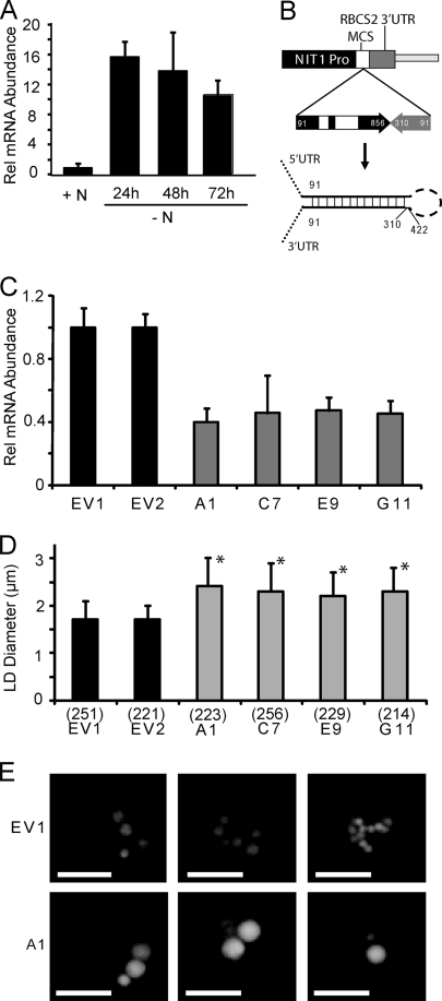
Eukaryotic cells store oils in the chemical form of triacylglycerols in distinct organelles, often called lipid droplets. These dynamic storage compartments have been intensely studied in the context of human health and also in plants as a source of vegetable oils for human consumption and for chemical or biofuel feedstocks. Many microalgae accumulate oils, particularly under conditions limiting to growth, and thus have gained renewed attention as a potentially sustainable feedstock for biofuel production. However, little is currently known at the cellular or molecular levels with regard to oil accumulation in microalgae, and the structural proteins and enzymes involved in the biogenesis, maintenance, and degradation of algal oil storage compartments are not well studied. Focusing on the model green alga Chlamydomonas reinhardtii, the accumulation of triacylglycerols and the formation of lipid droplets during nitrogen deprivation were investigated. Mass spectrometry identified 259 proteins in a lipid droplet-enriched fraction, among them a major protein, tentatively designated major lipid droplet protein (MLDP). This protein is specific to the green algal lineage of photosynthetic organisms. Repression of MLDP gene expression using an RNA interference approach led to increased lipid droplet size, but no change in triacylglycerol content or metabolism was observed.


Figures

FIG. 1.
FIG. 1.
Accumulation of TAGs in lipid droplets during N limitation. (A) Quantification of total cellular fatty acids (black bars) and fatty acids in TAG (gray bars) of dw15 cells grown in medium with 10 mM NH4+ (+N), or cells switched (arrow) to medium with no NH4+ (−N) and grown for 24, 48, or 72 h. (B) Confocal microscopy images of Nile red-stained CC-125 cells grown in medium with 10 mM NH4+ or cells switched (arrow) to medium with 0 mM NH4+ and grown for 12, 24, or 48 h. Differential interference contrast (D), chlorophyll autofluorescence (C), Nile red fluorescence indicating lipid droplets (L), and all three images merged (M) are shown. The scale bars indicate 5 μm.
FIG. 2.
FIG. 2.
Ultrastructure of cells during N limitation. Electron micrographs of a representative CC-125 cell grown in medium containing 10 mM NH4+ (0d) and representative cells switched to 0 mM NH4+ and grown for 1 (1d), 2 (2d), or 3 (3d) days, respectively, are shown. Scale bars represent 2 μm. E, eyespot; LD, lipid droplets; N, nucleus; P, pyranoid; S, starch granules; T, thylakoid membranes; V, vacuoles.
FIG. 3.
FIG. 3.
Characteristics of isolated lipid droplets. (A) Absorbance spectra of ethanol extracts (96%) normalized on an equal fatty acid basis from thylakoids of cells grown in medium containing 10 mM NH4+ (+N) or cells switched to 0 mM NH4+ and grown for 1 day (−N 1d) from enriched eyespot fraction and from lipid droplets were recorded. (B) Ratios of fatty acids derived from TAG (TAG FA) to total fatty acids (Total FA) for extracts from whole cells grown in medium with 10 mM NH4+ (+N) or whole cells switched to and grown in medium with no NH4+ (−N) for 24, 48, or 72 h, the enriched eyespot fraction (Eye), and isolated lipid droplets (LD) as indicated. (C) Proteins (30 μg) separated by gradient (4 to 20%) SDS-PAGE from whole cells (Tot. Prot., total protein), thylakoids, eyespots, and lipid droplets as indicated. The positions of molecular mass markers in kDa are indicated on the left, and sections excised from the gel separating lipid droplet-associated protein are indicated on the right.
FIG. 4.
FIG. 4.
Induction and RNAi suppression of MLDP gene expression. (A) Relative abundance of MLDP mRNA normalized to RACK1 (CBLP) in wild-type dw15 cells grown in medium with 10 mM NH4+ (+N), or dw15 cells switched to and grown in medium with no NH4+ (−N) for 24, 48, or 72 h measured by quantitative RT-PCR. Data are expressed as fold changes compared to the +N growth condition, which is given a value of 1. The standard deviation of three biological repeats, each done in triplicate, is shown. (B) Diagram showing the MLDP-RNAi construct designed with the NIT1 inducible promoter (Pro) of vector pNITPRO1 driving the expression of an MLDP genomic inverted repeat (numbers indicate base pairs from predicted 5′ UTR), and the predicted transcribed RNA hairpin product. (C) QRT-PCR analysis of MLDP mRNA levels in two empty vector control lines (EV1 and EV2), and four MLDP-RNAi lines isolated from four independent transformation events (A1, C7, E9, G11). Data are represented as percent change compared to EV1 and normalized to RACK1 as in panel A. Three replicates were averaged and standard deviation is shown. (D) Analysis of apparent lipid droplet diameter in empty vector controls and MLDP-RNAi lines described in (C). The average and standard deviation are shown for n (indicated in parentheses below each bar) lipid droplets counted, and cell lines are marked with an asterisk, for which P < 0.01 compared to EV1 in a two-tailed Student t test. (E) Fluorescent micrographs of representative cells of lines EV1 and A1 stained for lipid droplets with Nile red.
FIG. 5.
FIG. 5.
Northern blot analysis of MLDP. Five micrograms of total RNA isolated from cells grown in 10 mM NH4Cl (10 mM N) and cells deprived of N for 2 days (−N 2 d) was processed for Northern blotting as previously described (23). MLDP mRNA was specifically detected using a 32P-radiolabeled probe amplified from cDNA with forward and reverse primers 5′-ATGGTGGTGATCTTGTTTGACAGAG-3′ and 5′-CATTCCGCTGTACCTCCAGG-3′. Total RNA was stained with methylene blue (23) and shown as a loading control (rRNA). The results for two biological replicates of this experiment are shown (panels 1 and 2).
FIG. 6.
FIG. 6.
Multiple sequence alignment of MLDP with other green algal orthologs and hydropathy plots of MLDP and other known lipid droplet binding proteins. (A) MLDP and putative orthologs identified in the Chlorella sp. NC64A (http://genome.jgi-psf.org/ChlNC64A_1/ChlNC64A_1.home.html), Chlorella vulgaris C-169 (http://genome.jgi-psf.org/Chlvu1/Chlvu1.home.html), and Volvox carteri (http://genome.jgi-psf.org/Volca1/Volca1.home.html) genomes. Conserved residues are indicated with asterisks, and the coloring indicates the CORE index, where red and blue indicate the highest and lowest probability of the alignment being correct at a given residue, respectively, with colors in between the spectrum of red and blue representing a range of intermediate probabilities. Hydropathy plots for the proteins (followed by NCBI accession numbers in parentheses) MLDP (XP_001697668) (B), mouse ADRP (NP_031434) (C), mouse perilipin (NP_783571) (D), and Arabidopsis OLE1 (NP_194244) (E).

References

    1. Abramoff, M. D., P. J. Magelhaes, and S. J. Ram. 2004. Image processing with ImageJ. Biophotonics Int. 11:36-42.
    1. Allen, M. D., J. Kropat, S. Tottey, J. A. Del Campo, and S. S. Merchant. 2007. Manganese deficiency in Chlamydomonas results in loss of photosystem II and MnSOD function, sensitivity to peroxides, and secondary phosphorus and iron deficiency. Plant Physiol. 143:263-277. - PMC - PubMed
    1. Altschul, S. F., T. L. Madden, A. A. Schaffer, J. Zhang, Z. Zhang, W. Miller, and D. J. Lipman. 1997. Gapped BLAST and PSI-BLAST: a new generation of protein database search programs. Nucleic Acids Res. 25:3389-3402. - PMC - PubMed
    1. Athenstaedt, K., D. Zweytick, A. Jandrositz, S. D. Kohlwein, and G. Daum. 1999. Identification and characterization of major lipid particle proteins of the yeast Saccharomyces cerevisiae. J. Bacteriol. 181:6441-6448. - PMC - PubMed
    1. Atteia, A., A. Adrait, S. Brugiere, M. Tardif, L. R. van, O. Deusch, T. Dagan, L. Kuhn, B. Gontero, W. Martin, J. Garin, J. Joyard, and N. Rolland. 2009. A proteomic survey of Chlamydomonas reinhardtii mitochondria sheds new light on the metabolic plasticity of the organelle and on the nature of the alpha-proteobacterial mitochondrial ancestor. Mol. Biol. Evol. 26:1533-1548. - PubMed

Publication types

LinkOut - more resources