

Inducible costimulator promotes helper T-cell differentiation through phosphoinositide 3-kinase
- PMID: 19915142
- PMCID: PMC2787139
- DOI: 10.1073/pnas.0911573106
Inducible costimulator promotes helper T-cell differentiation through phosphoinositide 3-kinase
Abstract
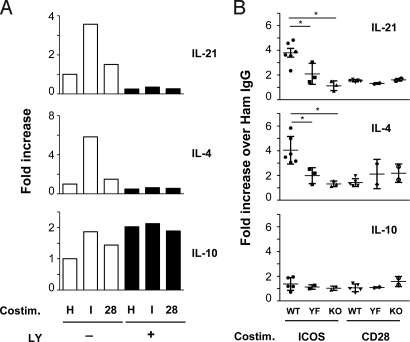
The T-cell costimulatory receptors, CD28 and the inducible costimulator (ICOS), are required for the generation of follicular B helper T cells (T(FH)) and germinal center (GC) reaction. A common signal transducer used by CD28 and ICOS is the phosphoinositide 3-kinase (PI3K). Although it is known that CD28-mediated PI3K activation is dispensable for GC reaction, the role of ICOS-driven PI3K signaling has not been defined. We show here that knock-in mice that selectively lost the ability to activate PI3K through ICOS had severe defects in T(FH) generation, GC reaction, antibody class switch, and antibody affinity maturation. In preactivated CD4(+) T cells, ICOS delivered a potent PI3K signal that was critical for the induction of the key T(FH) cytokines, IL-21 and IL-4. Under the same settings, CD28 was unable to activate PI3K but supported a robust secondary expansion of T cells. Thus, our results demonstrate a nonredundant function of ICOS-PI3K pathway in the generation of T(FH) and suggest that CD28 and ICOS play differential roles during a multistep process of T(FH) differentiation.
Conflict of interest statement
The authors declare no conflict of interest.
Figures
Similar articles
-
CD28 and inducible costimulatory protein Src homology 2 binding domains show distinct regulation of phosphatidylinositol 3-kinase, Bcl-xL, and IL-2 expression in primary human CD4 T lymphocytes.J Immunol. 2003 Jul 1;171(1):166-74. doi: 10.4049/jimmunol.171.1.166. J Immunol. 2003. PMID: 12816995
-
The inducible costimulator (ICOS) is critical for the development of human T(H)17 cells.Sci Transl Med. 2010 Oct 27;2(55):55ra78. doi: 10.1126/scitranslmed.3000448. Sci Transl Med. 2010. PMID: 20980695 Free PMC article.
-
Inducible costimulator facilitates T-dependent B cell activation by augmenting IL-4 translation.Mol Immunol. 2014 May;59(1):46-54. doi: 10.1016/j.molimm.2014.01.008. Epub 2014 Jan 31. Mol Immunol. 2014. PMID: 24486724
-
CD28 and CTLA-4 coreceptor expression and signal transduction.Immunol Rev. 2009 May;229(1):12-26. doi: 10.1111/j.1600-065X.2009.00770.x. Immunol Rev. 2009. PMID: 19426212 Free PMC article. Review.
-
Inducible T-cell co-stimulator: Signaling mechanisms in T follicular helper cells and beyond.Immunol Rev. 2019 Sep;291(1):91-103. doi: 10.1111/imr.12771. Immunol Rev. 2019. PMID: 31402504 Review.
Cited by
-
Follicular T-helper cell recruitment governed by bystander B cells and ICOS-driven motility.Nature. 2013 Apr 25;496(7446):523-7. doi: 10.1038/nature12058. Nature. 2013. PMID: 23619696
-
ICOS maintains the T follicular helper cell phenotype by down-regulating Krüppel-like factor 2.J Exp Med. 2015 Feb 9;212(2):217-33. doi: 10.1084/jem.20141432. Epub 2015 Feb 2. J Exp Med. 2015. PMID: 25646266 Free PMC article.
-
ICOS Co-Stimulation: Friend or Foe?Front Immunol. 2016 Aug 10;7:304. doi: 10.3389/fimmu.2016.00304. eCollection 2016. Front Immunol. 2016. PMID: 27559335 Free PMC article. Review.
-
Anti-chlamydial Th17 responses are controlled by the inducible costimulator partially through phosphoinositide 3-kinase signaling.PLoS One. 2012;7(12):e52657. doi: 10.1371/journal.pone.0052657. Epub 2012 Dec 20. PLoS One. 2012. PMID: 23285133 Free PMC article.
-
CXCR5 and ICOS expression identifies a CD8 T-cell subset with TFH features in Hodgkin lymphomas.Blood Adv. 2018 Aug 14;2(15):1889-1900. doi: 10.1182/bloodadvances.2018017244. Blood Adv. 2018. PMID: 30087107 Free PMC article.
References
Publication types
MeSH terms
Substances
Grants and funding
LinkOut - more resources
Full Text Sources
Other Literature Sources
Molecular Biology Databases
Research Materials
Miscellaneous
