

Role of hypoxia-inducible factor 1{alpha} in modulating cobalt-induced lung inflammation
- PMID: 19915160
- PMCID: PMC2822559
- DOI: 10.1152/ajplung.00252.2009
Role of hypoxia-inducible factor 1{alpha} in modulating cobalt-induced lung inflammation
Abstract
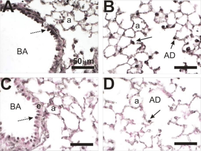
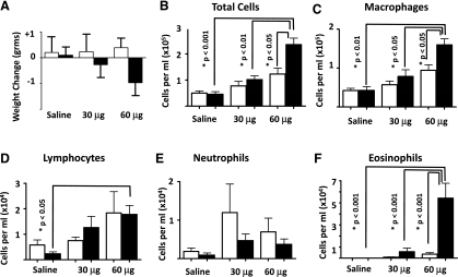
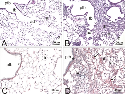
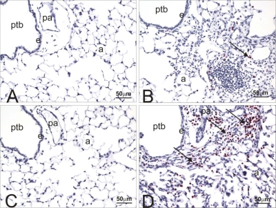
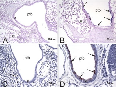
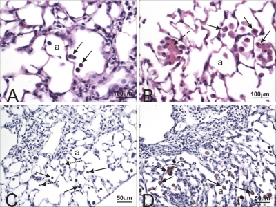
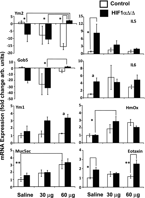
Hypoxia plays an important role in development, cellular homeostasis, and pathological conditions, such as cancer and stroke. There is also growing evidence that hypoxia is an important modulator of the inflammatory process. Hypoxia-inducible factors (HIFs) are a family of proteins that regulate the cellular response to oxygen deficit, and loss of HIFs impairs inflammatory cell function. There is little known, however, about the role of epithelial-derived HIF signaling in modulating inflammation. Cobalt is capable of eliciting an allergic response and promoting HIF signaling. To characterize the inflammatory function of epithelial-derived HIF in response to inhaled cobalt, a conditional lung-specific HIF1alpha, the most ubiquitously expressed HIF, deletion mouse, was created. Control mice showed classic signs of metal-induced injury following cobalt exposure, including fibrosis and neutrophil infiltration. In contrast, HIF1alpha-deficient mice displayed a Th2 response that resembled asthma, including increased eosinophilic infiltration, mucus cell metaplasia, and chitinase-like protein expression. The results suggest that epithelial-derived HIF signaling has a critical role in establishing a tissue's inflammatory response, and compromised HIF1alpha signaling biases the tissue towards a Th2-mediated reaction.
Figures
Similar articles
-
Acute cobalt-induced lung injury and the role of hypoxia-inducible factor 1alpha in modulating inflammation.Toxicol Sci. 2010 Aug;116(2):673-81. doi: 10.1093/toxsci/kfq155. Epub 2010 May 28. Toxicol Sci. 2010. PMID: 20511350 Free PMC article.
-
Loss of hypoxia-inducible factor 2 alpha in the lung alveolar epithelium of mice leads to enhanced eosinophilic inflammation in cobalt-induced lung injury.Toxicol Sci. 2014 Feb;137(2):447-57. doi: 10.1093/toxsci/kft253. Epub 2013 Nov 11. Toxicol Sci. 2014. PMID: 24218148 Free PMC article.
-
Neonatal epithelial hypoxia inducible factor-1α expression regulates the response of the lung to experimental asthma.Am J Physiol Lung Cell Mol Physiol. 2012 Mar 1;302(5):L455-62. doi: 10.1152/ajplung.00193.2011. Epub 2011 Dec 16. Am J Physiol Lung Cell Mol Physiol. 2012. PMID: 22180657 Free PMC article.
-
HIF-1α is a key mediator of the lung inflammatory potential of lithium-ion battery particles.Part Fibre Toxicol. 2019 Sep 18;16(1):35. doi: 10.1186/s12989-019-0319-z. Part Fibre Toxicol. 2019. PMID: 31533843 Free PMC article.
-
Mechanisms Leading to Differential Hypoxia-Inducible Factor Signaling in the Diabetic Kidney: Modulation by SGLT2 Inhibitors and Hypoxia Mimetics.Am J Kidney Dis. 2021 Feb;77(2):280-286. doi: 10.1053/j.ajkd.2020.04.016. Epub 2020 Jul 23. Am J Kidney Dis. 2021. PMID: 32711072 Review.
Cited by
-
Acute cobalt-induced lung injury and the role of hypoxia-inducible factor 1alpha in modulating inflammation.Toxicol Sci. 2010 Aug;116(2):673-81. doi: 10.1093/toxsci/kfq155. Epub 2010 May 28. Toxicol Sci. 2010. PMID: 20511350 Free PMC article.
-
Loss of Hif-2α Rescues the Hif-1α Deletion Phenotype of Neonatal Respiratory Distress In Mice.PLoS One. 2015 Sep 30;10(9):e0139270. doi: 10.1371/journal.pone.0139270. eCollection 2015. PLoS One. 2015. PMID: 26422241 Free PMC article.
-
Contributions of nonhematopoietic cells and mediators to immune responses: implications for immunotoxicology.Toxicol Sci. 2015 Jun;145(2):214-32. doi: 10.1093/toxsci/kfv060. Toxicol Sci. 2015. PMID: 26008184 Free PMC article. Review.
-
Activation of hypoxia-inducible factor-1 protects airway epithelium against oxidant-induced barrier dysfunction.Am J Physiol Lung Cell Mol Physiol. 2011 Dec;301(6):L993-L1002. doi: 10.1152/ajplung.00250.2011. Epub 2011 Sep 16. Am J Physiol Lung Cell Mol Physiol. 2011. PMID: 21926263 Free PMC article.
-
Klebsiella pneumoniae Siderophores Induce Inflammation, Bacterial Dissemination, and HIF-1α Stabilization during Pneumonia.mBio. 2016 Sep 13;7(5):e01397-16. doi: 10.1128/mBio.01397-16. mBio. 2016. PMID: 27624128 Free PMC article.
References
-
- Bucher JR, Elwell MR, Thompson MB, Chou BJ, Renne R, Ragan HA. Inhalation toxicity studies of cobalt sulfate in F344/N rats and B6C3F1 mice. Fundam Appl Toxicol 15: 357–372, 1990 - PubMed
-
- Bucher JR, Hailey JR, Roycroft JR, Haseman JK, Sills RC, Grumbein SL, Mellick PW, Chou BJ. Inhalation toxicity and carcinogenicity studies of cobalt sulfate. Toxicol Sci 49: 56–67, 1999 - PubMed
-
- Bunn HF, Poyton RO. Oxygen sensing and molecular adaptation to hypoxia. Phys Rev 76: 839–885, 1996 - PubMed
-
- Chupp GL, Lee CG, Jarjour N, Shim YM, Holm CT, He S, Dziura JD, Reed J, Coyle AJ, Kiener P, Cullen M, Grandsaigne M, Dombret MC, Aubier M, Pretolani M, Elias JA. A chitinase-like protein in the lung and circulation of patients with severe asthma. N Engl J Med 357: 2016–2027, 2007 - PubMed
Publication types
MeSH terms
Substances
Grants and funding
LinkOut - more resources
Full Text Sources
Molecular Biology Databases