. 2009 Dec;68(12):1294-308.
doi: 10.1097/NEN.0b013e3181c34bbe.

Chronic spinal injury repair by olfactory bulb ensheathing glia and feasibility for autologous therapy

Affiliations

Chronic spinal injury repair by olfactory bulb ensheathing glia and feasibility for autologous therapy

Cintia Muñoz-Quiles et al. J Neuropathol Exp Neurol. 2009 Dec.

Abstract

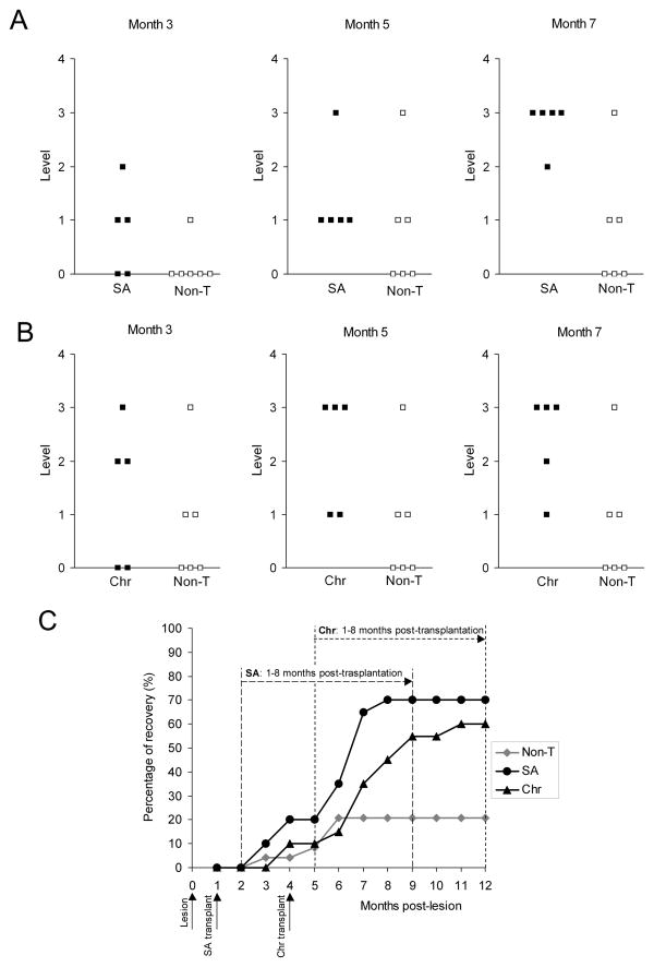
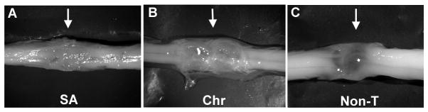
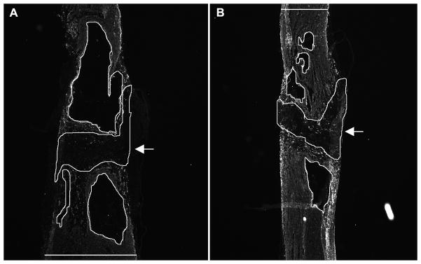
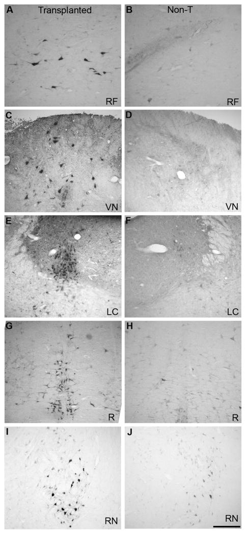
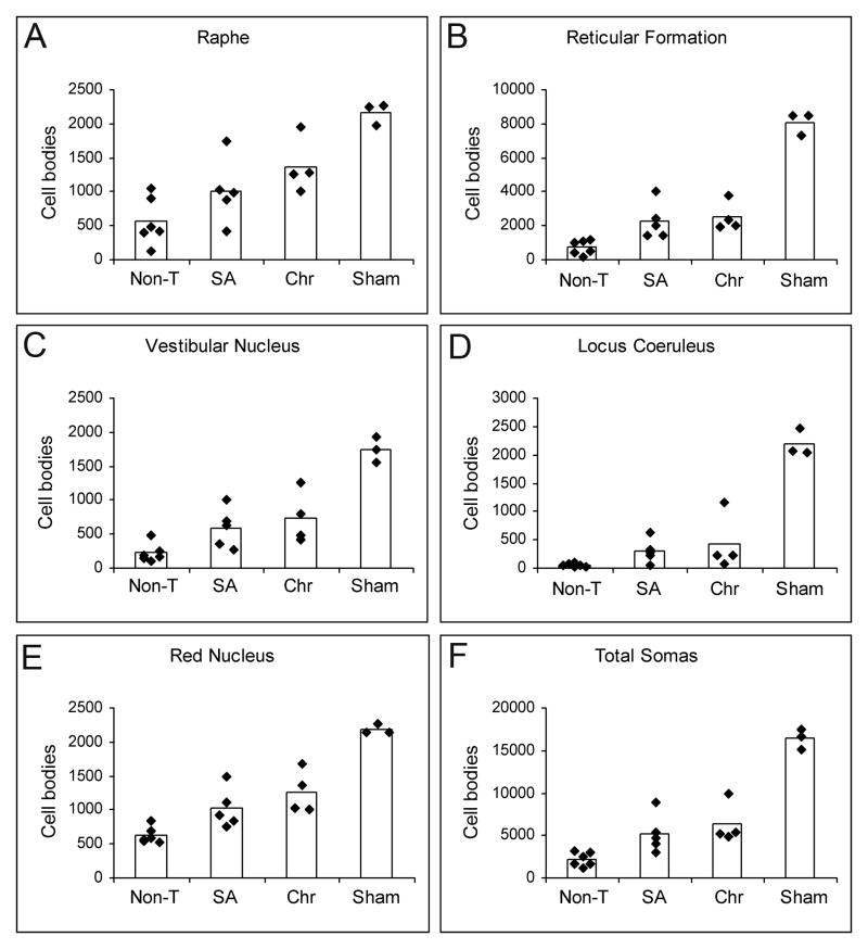
Olfactory bulb ensheathing glia (OB-OEG) promote repair of spinal cord injury (SCI) in rats after transplantation at acute or subacute (up to 45 days) stages. The most relevant clinical scenario in humans, however, is chronic SCI, in which no more major cellular or molecular changes occur at the injury site; this occurs after the third month in rodents. Whether adult OB-OEG grafts promote repair of severe chronic SCI has not been previously addressed. Rats with complete SCI that were transplanted with OB-OEG 4 months after injury exhibited progressive improvement in motor function and axonal regeneration from different brainstem nuclei across and beyond the SCI site. A positive correlation between motor outcome and axonal regeneration suggested a role for brainstem neurons in the recovery. Functional and histological outcomes did not differ after transplantation at subacute or chronic stages. Thus, autologous transplantation is a feasible approach as there is a time frame for patient stabilization and OEG preparation; moreover, the healing effects of OB-OEG on established injuries may offer new therapeutic opportunities for chronic SCI patients.


Figures

Figure 1
Figure 1
Appearance of the surgical field during different phases of the procedure. (A) After spinal cord transection, both stumps were lifted to ensure completeness of the lesion. (B) Spinal cord stumps were placed back into the vertebral channel apposing one another. (C–F) Lesion region during the second surgery (access to the spinal cord for transplantation). (C) The resin bridge (arrow) is firmly sealed to the spinous processes and laminae of adjacent intact vertebrae. (D) After bridge removal, a dense layer of connective tissue (arrowheads) was removed to expose the underlying spinal cord (arrow). (E) Dorsal aspect of both spinal cord stumps 4 months after complete transection. (F) Transplantation of olfactory bulb-ensheathing glia (OB-OEG) using a glass micro-needle (arrow). Inset: a detail of an OB-OEG culture used for transplantation and immunolabeled with anti-p75. Arrows in A, B and E indicate the transection sites.
Figure 2
Figure 2
Video frames of a paraplegic rat transplanted with olfactory bulb-ensheathing glia at the chronic stage, achieving the third climbing level, 5 months after transplantation. Detail of the hind limb movement indicates that this rat supports its weight and propels its body to reach the horizontal platform. The video of this animal is part of Video, Supplemental Digital Content 1, http://links.lww.com/NEN/A62.
Figure 3
Figure 3
Functional recovery of paraplegic rats after olfactory bulb-ensheathing glia transplantation at subacute (SA) and chronic (Chr) stages. (A, B) Levels achieved in the climbing test by (A) non-transplanted (Non-T) and SA, and by (B) Non-T and Chr rats at 3, 5 and 7 months post-grafting. Transplanted rats showed a functional recovery significantly higher than Non-T (SA: p < 0.001 and Chr: p = 0.001). Differences started to be significant from month 6 in SA rats (month 6: p = 0.029, months 7 and 8: p = 0.020), and from month 7 in Chr (p = 0.045). There were no differences between SA and Chr groups (compare A and B). (C) Progression of the functional outcome in all groups from the first month post-lesion until the end of the survival period (month 12). Each point represents the percentage of functional recovery exhibited by animals of each group. Dashed lines show the periods used to compare the outcome of Non-T vs. SA (long dash) and Non-T vs. Chr animals (short dash) from the first to the eighth month post-grafting. Non-T animals (grey rhombi) showed a slight but insignificant recovery. Before grafting, Chr and SA groups behaved as Non-T animals. SA group (black circles) and Chr (black triangles) started improving 2 months after transplantation (3 and 6 months post-lesion, respectively). Improvement of SA and Chr rats commenced at the second month, and began to be significant from the fifth (p = 0.004) and fourth months (p = 0.021), respectively.
Figure 4
Figure 4
Transection sites of olfactory bulb-ensheathing glia-grafted and non-transplanted spinal cords 12 months after lesion. Representative macroscopic images from subacute (SA) (A) and chronic (Chr) (B) transplanted rats and from a non-transplanted (Non-T) rat. Rostral and caudal stumps in all transplanted cords were bridged by white, opaque tissue (A, B); cord stumps in non-T rats were joined by a translucent membrane (C).
Figure 5
Figure 5
Longitudinal spinal cord sections showing areas used for quantification of glial-fibrillary acidic protein (GFAP)-negative tissue at the injury site. Sections of non-transferred (A) and transplanted (B) spinal cords immunolabeled with anti-GFAP. Encircled lines represent both GFAP-negative fibrous scar (arrows) and cysts (without arrows). The widths of the cords were also measured in these sections (horizontal lines in A and B). Top: rostral; bottom: caudal.
Figure 6
Figure 6
Brainstem coronal sections showing peroxidase-traced neurons in olfactory bulb-ensheathing glia (OB-OEG)-transplanted (A, C, E, G, I) and non-transplanted (B, D, F, H, J) rats. (A, B) Reticular formation (RF). (C, D) Vestibular nucleus (VN). (E, F) Locus coeruleus (LC). (G, H) Raphe (R). (I, J) Red nucleus (RN). Scale bar: A–J = 50 μm. Top: dorsal; bottom: ventral.
Figure 7
Figure 7
Quantification of neurons regenerating axons beyond the injury region. (A–E) Histograms show the number of peroxidase-traced neurons in non-transplanted (Non-T), subacute (SA), chronic (Chr) and Sham groups counted in raphe (A), reticular formation (B), vestibular nuclei (C), locus coeruleus (D), red nuclei (E), and in all nuclei (F). Black rhombi represent the number of cell bodies per animal; bars are the average of cell bodies per group. The total number of traced neurons was significantly higher in SA and Chr animals than in Non-T, and also when comparing per nuclei (except for raphe in SA rats) (p < 0.05). There were not significant differences between SA and Chr groups in any case.

References

    1. Franssen EH, de Bree FM, Verhaagen J. Olfactory ensheathing glia: Their contribution to primary olfactory nervous system regeneration and their regenerative potential following transplantation into the injured spinal cord. Brain Res Rev. 2007;56:236–58. - PubMed
    1. Ramon-Cueto A, Nieto-Sampedro M. Regeneration into the spinal cord of transected dorsal root axons is promoted by ensheathing glia transplants. Exp Neurol. 1994;127:232–44. - PubMed
    1. Ramon-Cueto A, Plant GW, Avila J, et al. Long-distance axonal regeneration in the transected adult rat spinal cord is promoted by olfactory ensheathing glia transplants. J Neurosci. 1998;18:3803–15. - PMC - PubMed
    1. Ramon-Cueto A, Cordero MI, Santos-Benito FF, et al. Functional recovery of paraplegic rats and motor axon regeneration in their spinal cords by olfactory ensheathing glia. Neuron. 2000;25:425–35. - PubMed
    1. Imaizumi T, Lankford KL, Waxman SG, et al. Transplanted olfactory ensheathing cells remyelinate and enhance axonal conduction in the demyelinated dorsal columns of the rat spinal cord. J Neurosci. 1998;18:6176–85. - PMC - PubMed

Publication types