. 2009 Dec;68(12):1282-93.
doi: 10.1097/NEN.0b013e3181c1da10.

Caspase-6 activation in familial alzheimer disease brains carrying amyloid precursor protein or presenilin i or presenilin II mutations

Affiliations

Caspase-6 activation in familial alzheimer disease brains carrying amyloid precursor protein or presenilin i or presenilin II mutations

Steffen Albrecht et al. J Neuropathol Exp Neurol. 2009 Dec.

Erratum in

  • J Neuropathol Exp Neurol. 2010 Jan;69(1):110

Abstract

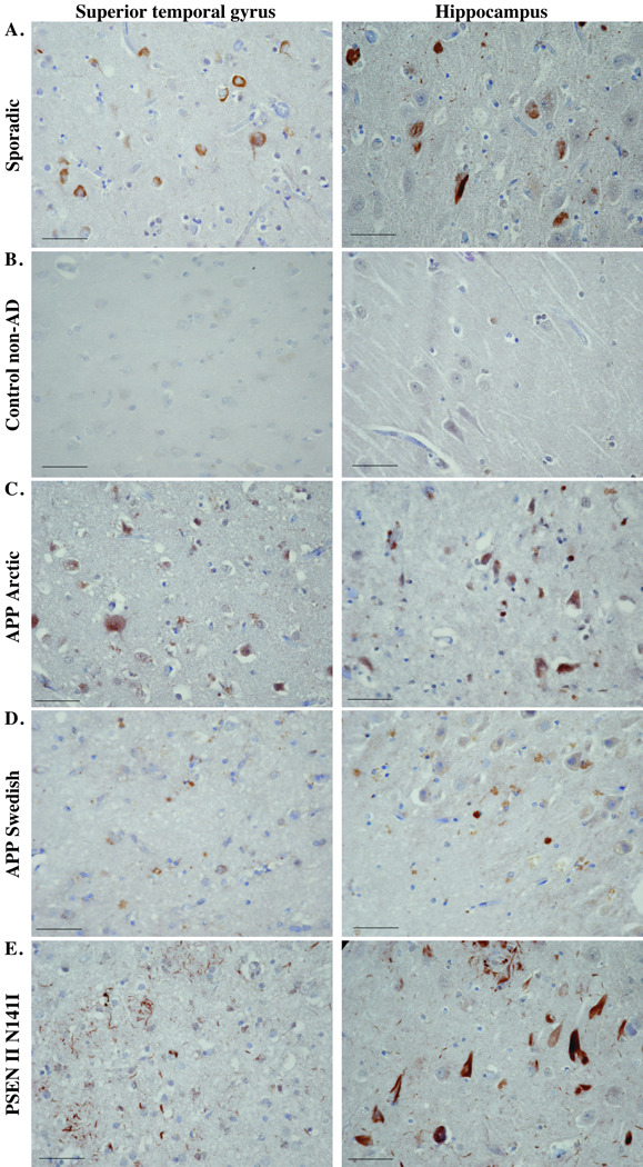
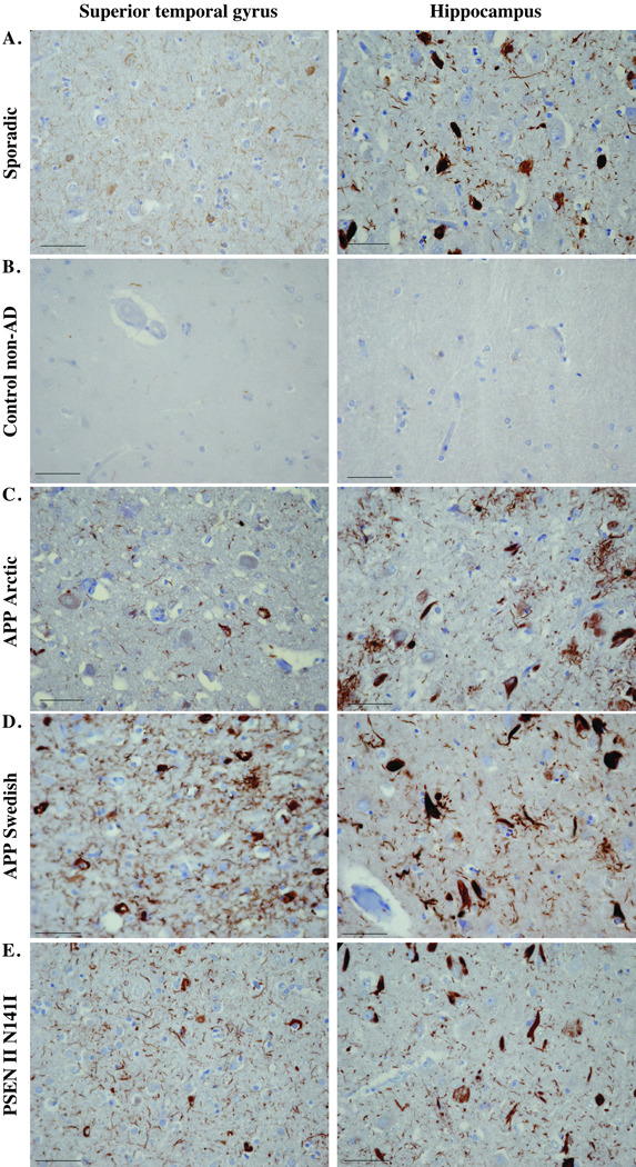
We previously demonstrated the activation of caspase-6 (Casp-6) in the hippocampus and cortex in cases of mild, moderate, severe, and very severe Alzheimer disease (AD). To determine whether Casp-6 is also activated in familial AD, we performed an immunohistochemical analysis of active Casp-6 and Tau cleaved by Casp-6 in temporal cortex and hippocampal tissue sections from cases of familial AD. The cases included 5 carrying the amyloid precursor protein K670N and M671L Swedish mutation, 1 carrying the amyloid precursor protein E693G Arctic mutation, 2 each carrying the Presenilin I M146V, F105L, A431E, V261F, and Y115C mutations, and 1 with the Presenilin II N141I mutation. Active Casp-6 immunoreactivity was found in all cases. Caspase-6 immunoreactivity was observed in neuritic plaques or in some cases cotton-wool plaques, and in neuropil threads and neurofibrillary tangles. These results indicate that Casp-6 is activated in familial forms of AD, as previously observed in sporadic forms. Because sporadic and familial AD cases have similar pathological features, these results support a fundamental role of Casp-6 in the pathophysiology of both familial and sporadic AD.


Figures

Figure 1
Figure 1
Immunohistochemical staining with 1277 anti-active Casp6 antiserum, anti-TauΔCasp6 antiserum, and anti-active Casp6 antiserum + anti-PHF-1 antibodies in the superior temporal gyrus (STG) and hippocampus of a representative familial Alzheimer disease (AD) case. Micrographs were taken with the 2x objective and reduced by 50% after importing the image. Detection of anti-active Casp6 and TauΔCasp6 was with diaminobenzidine; PHF-1 immunoreactivity was detected with Fast Red. The squares represent areas shown in Figures 2 and 3. Scale bars = 1 mm.
Figure 2
Figure 2
Immunohistochemical staining with anti-active Casp6 antiserum. Micrographs of anti-Casp6 immunostained superior temporal gyrus or hippocampus in the areas indicated in Figure 1 from a sporadic Alzheimer disease (AD) case (A), a non-AD case (B), an AD with the Arctic mutation (C), a representative AD APP Swedish case (D), an AD with the PSEN II N141I AD case (E), and a representative AD case of each of the PSEN I mutants indicated (FJ). Micrographs were taken with the 40x objective and reduced to 35%. Scale bars = 50 µm.
Figure 2
Figure 2
Immunohistochemical staining with anti-active Casp6 antiserum. Micrographs of anti-Casp6 immunostained superior temporal gyrus or hippocampus in the areas indicated in Figure 1 from a sporadic Alzheimer disease (AD) case (A), a non-AD case (B), an AD with the Arctic mutation (C), a representative AD APP Swedish case (D), an AD with the PSEN II N141I AD case (E), and a representative AD case of each of the PSEN I mutants indicated (FJ). Micrographs were taken with the 40x objective and reduced to 35%. Scale bars = 50 µm.
Figure 3
Figure 3
Immunohistochemical staining with 10635 anti-TauΔCasp6 antisera. Micrographs of TauΔCasp6 immunostained superior temporal gyrus or hippocampus in the areas indicated in Figure 1 from a sporadic Alzheimer disease (AD) case (A), a non-AD case (B), an AD with the Arctic mutation (C), a representative AD APP Swedish case (D), an AD with the PSEN II N141I AD case (E), and a representative AD case of each of the PSEN I mutants indicated (FJ). Micrographs were taken with the 40x objective and reduced to 35%. Scale bars = 50 µm.
Figure 3
Figure 3
Immunohistochemical staining with 10635 anti-TauΔCasp6 antisera. Micrographs of TauΔCasp6 immunostained superior temporal gyrus or hippocampus in the areas indicated in Figure 1 from a sporadic Alzheimer disease (AD) case (A), a non-AD case (B), an AD with the Arctic mutation (C), a representative AD APP Swedish case (D), an AD with the PSEN II N141I AD case (E), and a representative AD case of each of the PSEN I mutants indicated (FJ). Micrographs were taken with the 40x objective and reduced to 35%. Scale bars = 50 µm.
Figure 4
Figure 4
Co-immunostaining of active Casp6 with PHF-1. Micrographs show colocalization of active Casp6 (brown) and PHF-1 (red) in (A) neurofibrillary tangles (black arrow), (B) neuropil threads (black arrowhead) and a structure resembling a tangle stained only with anti-active Casp6 (white arrow). Left panel is a lower magnification indicating the area where the picture was taken (black arrow). Scale bar = 1 mm. Right panel is a higher magnification. Scale bar = 50 µm. (C) Micrograph showing punctate cytoplasmic anti-active Casp6 immunoreactivity in the APP Arctic case. The right panel represents a magnified image of the anti-active Casp6 immunopositive neurons with more or less PHF-1 immunoreactivity.

References

    1. Creagh EM, Conroy H, Martin SJ. Caspase-activation pathways in apoptosis and immunity. Immunol Rev. 2003;193:10–21. - PubMed
    1. Gervais F, Xu D, Robertson G, et al. Involvement of caspases in proteolytic cleavage of Alzheimer's ß-amyloid precursor protein and amyloidogenic ß-peptide formation. Cell. 1999;97:395–406. - PubMed
    1. LeBlanc AC, Liu H, Goodyer C, et al. Caspase-6 role in apoptosis of human neurons, amyloidogenesis and Alzheimer's disease. J Biol Chem. 1999;274:23426–23436. - PubMed
    1. Tesco G, Koh YH, Tanzi RE. Caspase activation increases beta-amyloid generation independently of caspase cleavage of the beta-amyloid precursor protein (APP) J Biol Chem. 2003;278:46074–46080. - PubMed
    1. Guo H, Albrecht S, Bourdeau M, et al. Active caspase-6 and caspase-6 cleaved tau in neuropil threads, neuritic plaques and neurofibrillary tangles of Alzheimer’s disease. Am J Pathol. 2004;165:523–531. - PMC - PubMed

Publication types

MeSH terms