

doi: 10.1038/nchembio.257.
Epub 2009 Nov 8.
An artificial di-iron oxo-protein with phenol oxidase activity
Affiliations
- PMID: 19915535
- PMCID: PMC3808167
- DOI: 10.1038/nchembio.257
Item in Clipboard
An artificial di-iron oxo-protein with phenol oxidase activity
Nat Chem Biol.
2009 Dec.
Abstract
Here we report the de novo design and NMR structure of a four-helical bundle di-iron protein with phenol oxidase activity. The introduction of the cofactor-binding and phenol-binding sites required the incorporation of residues that were detrimental to the free energy of folding of the protein. Sufficient stability was, however, obtained by optimizing the sequence of a loop distant from the active site.
Figures
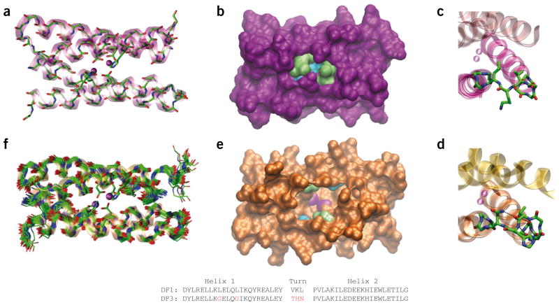
Comparison of DF1 and DF3 structures. (a) Di-Zn(II)-DF1 crystal structure. (b) Di-Zn(II)-DF1 surface representation, displaying the accessibility to the active site and highlighting the different residues in position 9 (lime) and 13 (cyan). (c) Di-Zn(II)-DF1 loop structure. (d) Di-Zn(II)-DF3 loop structure; N-capping and C-capping interactions are depicted. (e) Di-Zn(II)-DF3 surface representation, displaying the accessibility to the active site and highlighting the different residues in position 9 (lime) and 13 (cyan). The point mutations introduced in proximity of DF3 active site create a cavity large enough to allow substrates to approach the metal ions (depicted in magenta in e). In contrast, the active site is completely buried in DF1 (b). (f) NMR bundle of the best 30 minimized structures for di-Zn(II)-DF3. The structures were generated with Visual Molecular Dynamics (VMD; http://www.ks.uiuc.edu/Research/vmd/ ). DF1 and DF3 sequence comparison are also reported. Amino acid substitutions in the DF3 sequence are indicated in red. The atomic coordinates of di-Zn(II)-DF3 ensembles, chemical shifts and NMR constraints have been deposited in the Protein Data Bank under accession code 2KIK.
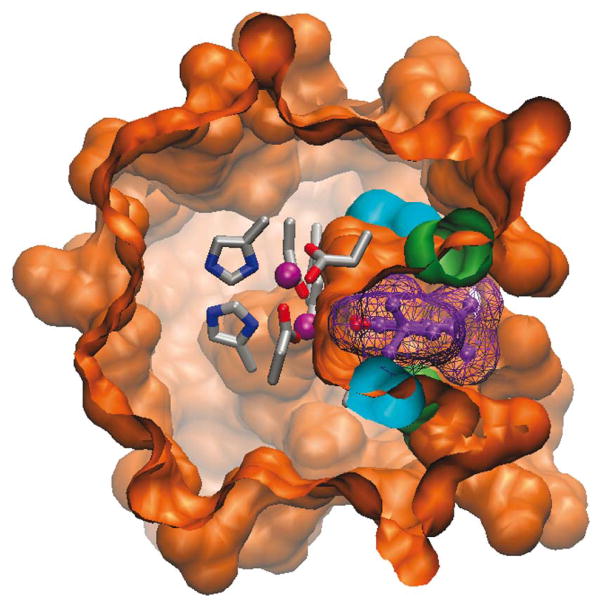
Model of substrate interaction with DF3 protein. Surface representation of the di-Zn(II)-DF3 NMR structure with a 3,5-DTBC molecule (violet) bound into the active site access channel. The surface associated with the bound 3,5-DTBC is also depicted. The model structure was generated with VMD.
References
Publication types
MeSH terms
Substances
Associated data
Grants and funding
LinkOut - more resources
Full Text Sources
Other Literature Sources
