

doi: 10.1038/nchembio.252.
Epub 2009 Nov 8.
Translating metabolic exchange with imaging mass spectrometry
Affiliations
- PMID: 19915536
- PMCID: PMC2778862
- DOI: 10.1038/nchembio.252
Item in Clipboard
Translating metabolic exchange with imaging mass spectrometry
Nat Chem Biol.
2009 Dec.
Abstract
Metabolic exchange between an organism and the environment, including interactions with neighboring organisms, is important for processes of organismal development. Here we develop and use thin-layer agar natural product MALDI-TOF imaging mass spectrometry of intact bacterial colonies grown on top of the MALDI target plate to study an interaction between two species of bacteria and provide direct evidence that Bacillus subtilis silences the defensive arsenal of Streptomyces coelicolor.
Figures
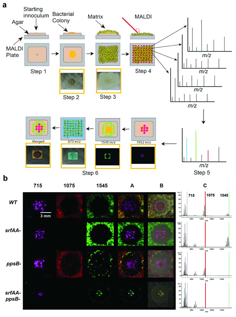
(a) Outline of the thin layer agar npMALDI-I on schematic of the thin layer agar npMALDI-I approach. Step 1, cover the MALDI plate with a thin layer of growth media and then inoculate with a small sample containing the bacteria. Step 2, allow the bacteria to grow. Step 3, cover the sample with matrix. Step 4, subject the sample to MALDI imaging. Step5, average all the spectra obtained in the imaging run. Step 6, display all the ions of interest as a color. (b) IMS of Bacillus subtilis 3610 and the srfAA, ppsB mutants and the ppsB and srfAA double mutant. A is the merged signal of m/z 715, 1075, and 1545. B is the merged signal with an optical image of the colony. The right side of this figure shows the average signal from m/z 600–1600 of all the spectra obtained in the imaging runs.
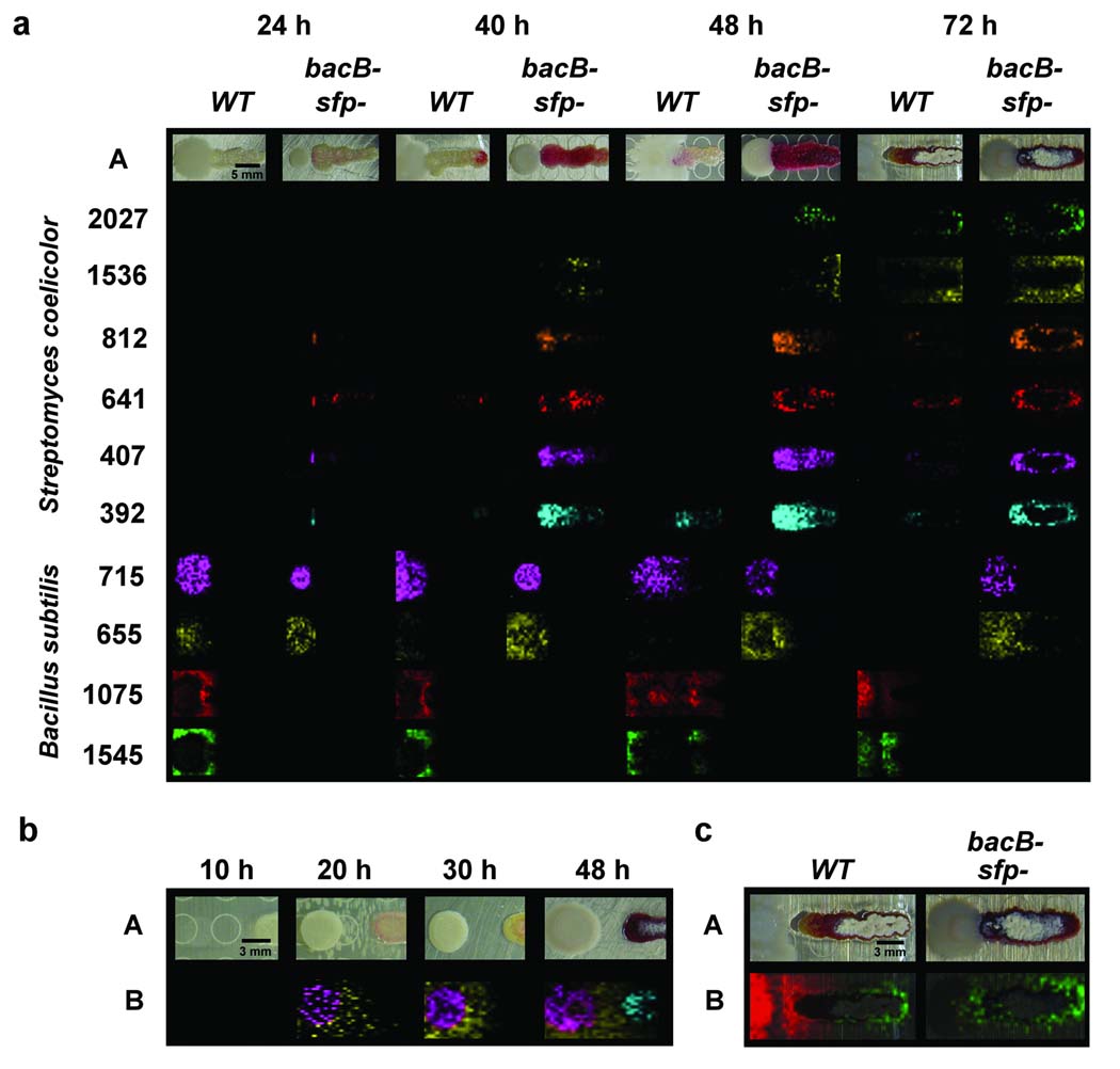
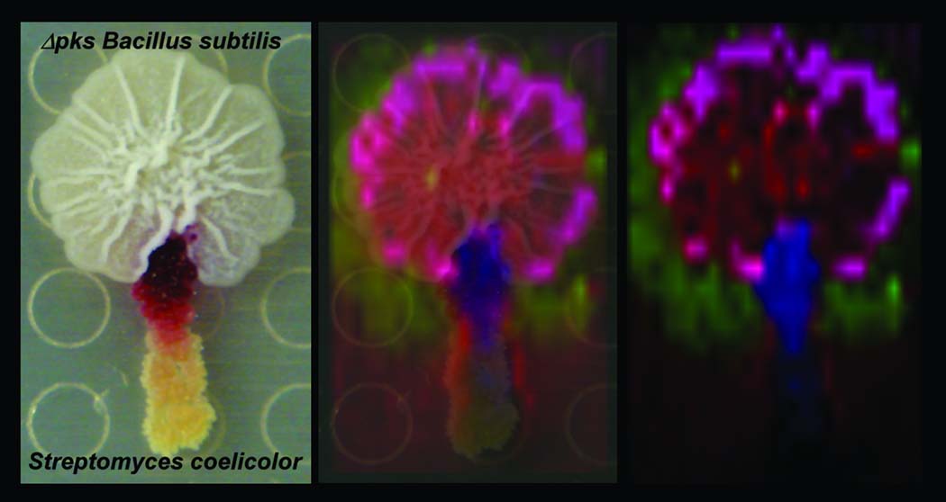
Time course IMS of Bacillus subtilis strains and Streptomyces coelicolor cohabitation. (a) Bacillus subtilis strains were spotted 5 mm away from Streptomyces coelicolor. A is optical photograph of colonies. m/z 392 is a representative prodiginines (6 and 7) [M+H]+. m/z 1075 is surfactin representative (2a–c) [M+K]+. m/z 1545 is plipastatin representative (3a–d) [M+K]+. m/z 1536 is CDA representative (10a–b) [M+H]+. m/z 2027 is sapB (9) [M+H]+. m/z 407, 641, 655 and 812 are unknown compounds. (b) bacB/sfp double deletion Bacillus subtilis strain were spotted 8 mm away from Streptomyces coelicolor. A is optical photograph of colonies. Purple is m/z 715. Yellow is the unknown ion at m/z 655. Blue are prodiginines (6 and 7). (c) The aerial hyphae formation and SapB (9) production by Streptomyces coelicolor is inhibited by surfactin (2a–c). A is an optical photograph of colonies. Red is surfactin (2a–c). Green is sapB (9).
References
Publication types
MeSH terms
Substances
Associated data
- PubChem-Substance/85244562
- PubChem-Substance/85244563
- PubChem-Substance/85244564
- PubChem-Substance/85244565
- PubChem-Substance/85244566
- PubChem-Substance/85244567
- PubChem-Substance/85244568
- PubChem-Substance/85244569
- PubChem-Substance/85244570
- PubChem-Substance/85244571
- PubChem-Substance/85244572
- PubChem-Substance/85244573
- PubChem-Substance/85244574
- PubChem-Substance/85244575
- PubChem-Substance/85244576
- PubChem-Substance/85244577
Grants and funding
LinkOut - more resources
Full Text Sources
Other Literature Sources
Molecular Biology Databases
