. 2009 Dec;5(12):936-46.
doi: 10.1038/nchembio.246. Epub 2009 Nov 1.

A synergistic small-molecule combination directly eradicates diverse prion strain structures

Affiliations

A synergistic small-molecule combination directly eradicates diverse prion strain structures

Blake E Roberts et al. Nat Chem Biol. 2009 Dec.

Abstract

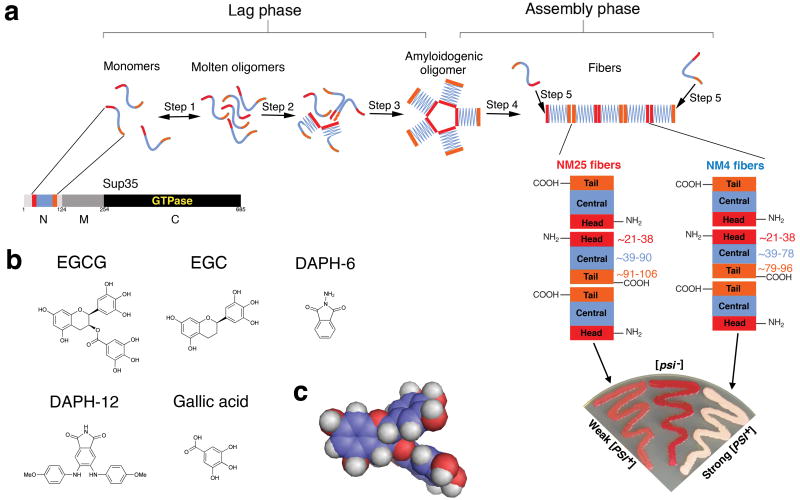
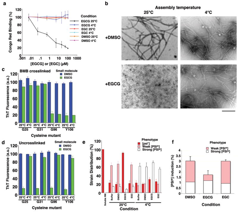
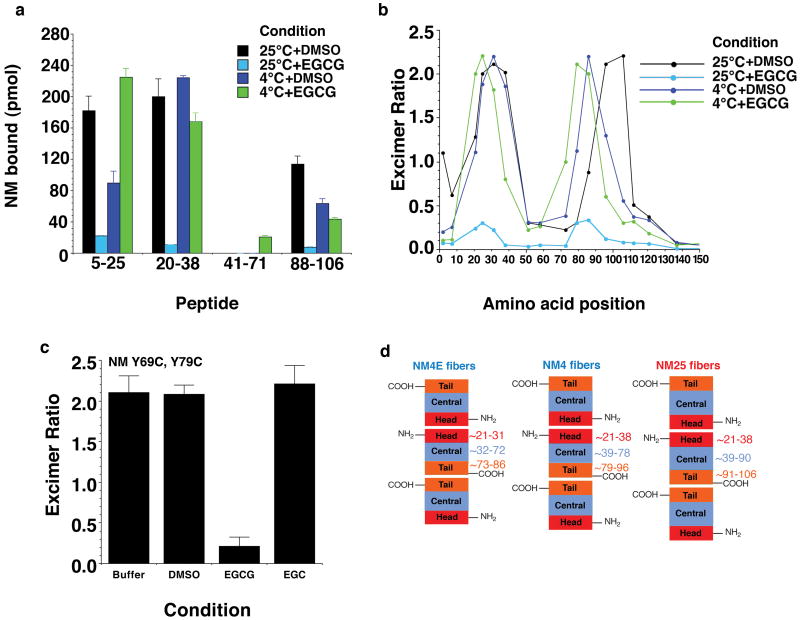
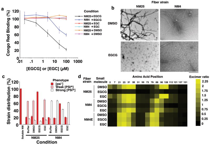
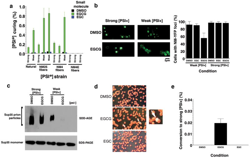
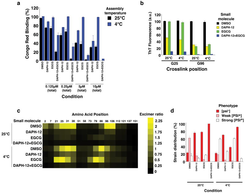
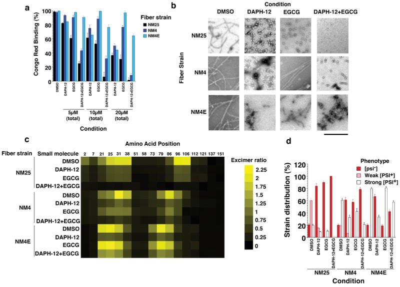
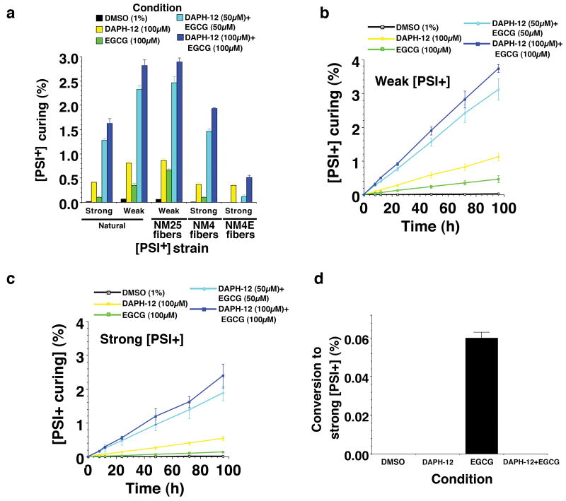
Safely eradicating prions, amyloids and preamyloid oligomers may ameliorate several fatal neurodegenerative disorders. Yet whether small-molecule drugs can directly antagonize the entire spectrum of distinct amyloid structures or 'strains' that underlie distinct disease states is unclear. Here, we investigated this issue using the yeast prion protein Sup35. We have established how epigallocatechin-3-gallate (EGCG) blocks synthetic Sup35 prionogenesis, eliminates preformed Sup35 prions and disrupts inter- and intramolecular prion contacts. Unexpectedly, these direct activities were strain selective, altered the repertoire of accessible infectious forms and facilitated emergence of a new prion strain that configured original, EGCG-resistant intermolecular contacts. In vivo, EGCG cured and prevented induction of susceptible, but not resistant strains, and elicited switching from susceptible to resistant forms. Importantly, 4,5-bis-(4-methoxyanilino)phthalimide directly antagonized EGCG-resistant prions and synergized with EGCG to eliminate diverse Sup35 prion strains. Thus, synergistic small-molecule combinations that directly eradicate complete strain repertoires likely hold considerable therapeutic potential.


Figures

Figure 1
Figure 1. Model of Sup35 prion assembly and chemical structure of EGCG
(a) Sup35 is composed of a C-terminal GTPase domain (amino acids 254-685, black) that confers translation termination activity, a highly charged middle domain (M, amino acids 124-253, dark grey), and a prionogenic N-terminal domain (N, amino acids 1-123, light grey) enriched in Gln, Asn, Tyr and Gly. Within N, prion recognition elements termed the ‘Head’ (red) and ‘Tail’ (orange), which flank a ‘Central Core’ (blue), are proposed to play important roles in prion formation,. (b) Mechanism of Sup35 prionogenesis in vitro. Only the Head (red), Central Core (blue), and Tail (orange) regions of N are depicted. During lag phase, an equilibrium is rapidly established between monomers (∼90% of total NM) and structurally labile, molten oligomers (∼10% of total NM) (step 1). The monomers within molten oligomers gradually rearrange (step 2) until they access the amyloidogenic oligomer conformation (step 3). This transient intermediate rapidly converts to the amyloid form at the end of lag phase (step 4). Amyloid fibers then assemble rapidly as they recruit and convert monomers at their ends (step 5). The prion recognition elements within N are proposed to make homotypic intermolecular contacts such that Sup35 prion fibers are held together by an alternating sequence of ‘Head-to-Head’ (red) and ‘Tail-to-Tail’ (orange) contacts. The Central Core (blue) is sequestered by intramolecular contacts. This schematic is not meant to imply precise details of molten oligomer or amyloidogenic oligomer structure. (c) Schematic of different Sup35 prion strain structures. NM25 fibers form after assembly at 25°C. NM25 fibers also form at 4°C when NM is chemically crosslinked, with BMB (a flexible 11Å crosslinker) in the Tail region (orange). NM4 fibers form after assembly at 4°C. NM4 fibers also form at 25°C when NM is chemically crosslinked with BMB in the Head region (red). Note that the Central Core (blue) and Tail (orange) are comprised of different amino acids in the NM25 and NM4 fiber conformations. We must note that the atomic structure of Sup35 prion strains remains uncertain and alternative models of Sup35 prion structure have been proposed,,,,,-. (d) Weak [PSI+] (pink), [psi-] (red) and strong [PSI+] (white) yeast growing on 25% YPD. These cells carry a premature stop codon in their ADE1 gene, do not make functional Ade1 and accumulate a red metabolite provided Sup35 is fully functional. Thus, colony color gives an indication of the extent of Sup35 aggregation and contingent loss-of-function. Color ranges from red in [psi-] colonies through pink in weak [PSI+] colonies to white in strong [PSI+] colonies as Sup35 aggregation and loss-of-function increases. Transformation of NM25 fibers into [psi-] cells yields mostly weak [PSI+], whereas transformation of NM4 fibers into [psi-] cells yields mostly strong [PSI+]. (e) Chemical structure of EGCG, DAPH-12, EGC, gallic acid, and DAPH-6. (f) Space-filling model of EGCG reveals a nonplanar structure.
Figure 2
Figure 2. EGCG inhibits assembly of select Sup35 prion strains
(a) Spontaneous, agitated NM (5μM) fibrillization after 4h at 25°C or 4°C in the presence of EGCG, EGC (0-100μM) or DMSO (0-1%). Fibrillization was measured by CR binding and 100% reflects assembly in the absence of EGCG, EGC or DMSO. Values represent means±SD (n=3-8). (b) NM was assembled as in (a) at 25°C or 4°C in the presence of DMSO (1%) or EGCG (20μM) and processed for EM. Bar, 0.5μm. (c, d) NM cysteine variants were crosslinked (c) under denaturing conditions with a flexible 11Å BMB crosslink at position 25, 31, 96 or 106. (d) Alternatively, NM cysteine variants were left uncrosslinked. The indicated NM protein (5μM) was then assembled with agitation at 25°C or 4°C for 4h in the presence of DMSO (1%) or EGCG (20μM). Fibrillization was measured by ThT fluorescence. Values represent means±SD (n=3). (e) NM (5μM) was incubated with agitation at 25°C or 4°C for 4h in the absence or presence of DMSO (1%), EGCG or EGC (20μM). Reactions were dialyzed to remove unbound small molecule, concentrated, sonicated and transformed into [psi-] cells. The proportion of [psi-], weak [PSI+] or strong [PSI+] transformants was then determined. Values represent means±SD (n=3). (f) NM-YFP was overexpressed in [psi-] Δpdr5 cells for 12h in the presence of DMSO (1%), EGCG or EGC (125μM). Cells were then plated on 25% YPD and the proportion of weak and strong [PSI+] colonies was determined. Values represent means±SD (n=3).
Figure 3
Figure 3. EGCG prevents inter- and intramolecular contact formation of select Sup35 prion strains
(a) The indicated N peptides were immobilized on nitrocellulose and incubated for 16h at either 25°C or 4°C with NM-his (1μM) in the presence of DMSO (1%) or EGCG (20μM). The amount of NM-his bound to each peptide was then determined by immunoblot using an anti-his antibody, followed by densitometry and comparison to known amounts of NM-his. Values represent means±SD (n=3). (b) Proximity analysis assessed by excimer fluorescence. NM proteins (5μM) carrying pyrene labels at the indicated single sites were incubated for 4h with agitation at 25°C or 4°C in the presence of DMSO (1%) or EGCG (20μM). The ratio of excimer fluorescence to non-excimer fluorescence (I465nm/I375nm) is plotted. Note that EGCG causes distinct intersubunit interfaces to form at 4°C and prevents their formation at 25°C. (c) NM (5μM) labeled with pyrene at positions 69 and 79 was incubated for 4h with agitation at 25°C in the absence or presence of DMSO (1%), EGCG or EGC (20μM). The ratio of excimer fluorescence to non-excimer fluorescence (I476nm/I384nm) is plotted. Values represent means±SD (n=3). (d) A new fiber strain, NM4E, forms in the presence of EGCG at 4°C. Note that the Head-to-Head contact and the Tail-to-Tail contact is more N-terminal relative to NM4 and NM25 fibers.
Figure 4
Figure 4. EGCG disrupts preformed inter- and intramolecular contacts of select Sup35 prion strains
(a) NM25 or NM4 fibers (2.5μM monomer) were incubated for 24h at 25°C with DMSO (1%), EGCG or EGC (0.025-100μM). Fiber integrity was then determined by CR binding. Values represent means±SD (n=3). (b) NM25 or NM4 fibers (2.5μM monomer) were incubated with DMSO (1%) or EGCG (20μM) for 24h and processed for EM. Bar, 0.5μm. (c) NM25 or NM4 fibers (2.5μM monomer) were incubated with or without DMSO (1%), EGCG or EGC (20μM) for 24h. Reactions were then dialyzed to remove unbound small molecule, concentrated, sonicated and transformed into [psi-] cells. The proportion of [psi-], weak [PSI+] or strong [PSI+] transformants was then determined. Values represent means±SD (n=3). (d) NM proteins (5μM) carrying pyrene labels at single sites were assembled into fibers by incubation for 16h with agitation at 25°C, 4°C or at 4°C in the presence of EGCG (20μM). Assembled NM25, NM4 or NM4E fibers (2.5μM monomer) were then treated with DMSO (1%), EGCG or EGC (25μM) for 24h. The ratio of excimer fluorescence to non-excimer fluorescence (I465nm/I375nm) is displayed.
Figure 5
Figure 5. EGCG eliminates select Sup35 prion strains
(a) We assessed EGCG curing of natural [PSI+] strain variants and several new [PSI+] strain variants generated by infecting [psi-] Δpdr5 cells with NM25 (two weak variants, one strong), NM4 (two strong variants, one weak) or NM4E (all strong). The indicated [PSI+] Δpdr5 strain variants were treated with DMSO (1%), EGCG or EGC (125μM) for 72h in liquid culture. Cells were plated on 25% YPD and the proportion of red [psi-] colonies was determined. Values represent means±SD (n=4). (b) NM-YFP was expressed for 5h in weak or strong [PSI+] Δpdr5 cells. Expression was then shut off in the presence of DMSO (1%), EGCG or EGC (250μM) for 2h, and cells were then imaged (left). Note the more diffuse fluorescence in weak [PSI+] cells treated with EGCG. The proportion of cells with NM-YFP foci was then determined (right). Values represent means±SD (n=16). (c) The indicated [PSI+] Δpdr5 strain variants were treated with either DMSO (1%) or EGCG (125μM) for 48h in liquid culture and then processed for SDD-AGE or SDS-PAGE followed by immunoblot to detect Sup35 prion particles or Sup35 monomers. (d, e) Weak [PSI+] Δpdr5 were treated with DMSO (1%), EGCG or EGC (125μM) for 48h in liquid culture. Cells were plated on 25% YPD (d). White arrows denote strong [PSI+] colonies that formed in the presence of EGCG, and one example is shown at higher magnification. The proportion of strong [PSI+] colonies was determined (e). Values represent means±SD (n=9).
Figure 6
Figure 6. Combinations of DAPH-12 and EGCG prevent formation of multiple prion strains
(a) Spontaneous, agitated NM (5μM) fibrillization after 6h at 25°C or 4°C in the presence of DMSO (1%), DAPH-12, EGCG or EGCG plus DAPH-12 (0.5:0.5). The total concentration of small molecule was kept constant at 0.125μM, 0.25μM, 5μM or 10μM. Fibrillization was measured by CR binding and 100% reflects assembly in the absence of DMSO, DAPH-12 or EGCG. Values represent means±SD (n=3). (b) NM cysteine variants were crosslinked under denaturing conditions with a flexible 11Å BMB crosslink at position 25 or 96. The indicated NM protein (5μM) was then assembled with agitation at 25°C or 4°C for 6h in the presence of DMSO (1%), DAPH-12 (10μM), EGCG (10μM) or DAPH-12 plus EGCG (5μM of each). Fibrillization was measured by ThT fluorescence. Values represent means±SD (n=3). (c) Proximity analysis assessed by excimer fluorescence. NM proteins (5μM) carrying pyrene labels at the indicated single sites were incubated for 6h with agitation at 25°C or 4°C in the presence of DMSO (1%), DAPH-12 (10μM), EGCG (10μM) or DAPH-12 plus EGCG (5μM of each). The ratio of excimer fluorescence to non-excimer fluorescence (I465nm/I375nm) is plotted. (d) NM (5μM) was incubated with agitation at 25°C or 4°C for 6h in the presence of DMSO (1%), DAPH-12 (10μM), EGCG (10μM) or DAPH-12 plus EGCG (5μM of each). Reactions were dialyzed to remove unbound small molecule, concentrated, sonicated and transformed into [psi-] cells. The proportion of [psi-], weak [PSI+] or strong [PSI+] transformants was then determined. Values represent means±SD (n=3).
Figure 7
Figure 7. Combinations of DAPH-12 and EGCG remodel multiple prion strains
(a) NM25, NM4 or NM4E fibers (2.5μM monomer) were incubated for 24h at 25°C with DMSO (1%), DAPH-12, EGCG or DAPH-12 plus EGCG (0.5:0.5). The total concentration of small molecule was kept constant at 5μM, 10μM or 20μM. Fiber integrity was then determined by CR binding. Values represent means±SD (n=3). (b) NM25, NM4 or NM4E fibers (2.5μM monomer) were incubated for 24h at 25°C with DMSO (1%), DAPH-12 (20μM), EGCG (20μM) or DAPH-12 plus EGCG (10μM of each). Reactions were then processed for EM. Bar, 0.5μm. (c) NM proteins (5μM) carrying pyrene labels at single sites were assembled into fibers by incubation for 16h with agitation at either 4°C, 25°C or 4°C in the presence of EGCG. Assembled fibers (2.5μM monomer) were then treated with DMSO (1%), DAPH-12 (20μM), EGCG (20μM) or DAPH-12 plus EGCG (10μM of each) for 24h. The ratio of excimer fluorescence to non-excimer fluorescence (I465nm/I375nm) is plotted. (d) NM25, NM4 or NM4E fibers (2.5μM monomer) were incubated with DMSO (1%), DAPH-12 (20μM), EGCG (20μM) or DAPH-12 plus EGCG (10μM of each) for 24h. Reactions were then dialyzed to remove unbound small molecule, concentrated, sonicated and transformed into [psi-] cells. The proportion of [psi-], weak [PSI+] or strong [PSI+] transformants was then determined. Values represent means±SD (n=3).
Figure 8
Figure 8. EGCG and DAPH-12 synergize to cure various [PSI+] variants
(a) The indicated [PSI+] Δpdr5 strain variants were treated with DMSO (1%), DAPH-12 (100μM), EGCG (100μM) or EGCG plus DAPH-12 (50μM of each or 100μM of each) for 72h in liquid culture. Cells were plated and the proportion of red [psi-] colonies was determined. Values represent means±SD (n=4). (b, c) Weak [PSI+] Δpdr5 cells (b) or strong [PSI+] Δpdr5 cells (c) were treated with DMSO (1%), DAPH-12 (100μM), EGCG (100μM) or EGCG plus DAPH-12 (50μM of each or 100μM of each) for 0-96h in liquid culture. At the indicated times, cells were plated and the proportion of red [psi-] colonies was determined. Values represent means±SD (n=3). (d) Weak [PSI+] Δpdr5 were treated with DMSO (1%), DAPH-12 (100μM), EGCG (100μM) or DAPH-12 plus EGCG (100μM of each) for 72h in liquid culture. Cells were plated and the proportion of strong [PSI+] colonies was determined. Values represent means±SD (n=5).

References

    1. Nelson R, Eisenberg D. Structural models of amyloid-like fibrils. Adv Protein Chem. 2006;73:235–82. - PubMed
    1. Skovronsky DM, Lee VMY, Trojanowski JQ. Neurodegenerative Diseases: New Concepts of Pathogenesis and Their Therapeutic Implications. Annu Rev Pathol Mech Dis. 2006;1:151–170. - PubMed
    1. Prusiner SB. Prions. Proc Natl Acad Sci U S A. 1998;95:13363–83. - PMC - PubMed
    1. Roberts BE, Shorter J. Escaping amyloid fate. Nat Struct Mol Biol. 2008;15:544–6. - PubMed
    1. Wells JA, McClendon CL. Reaching for high-hanging fruit in drug discovery at protein-protein interfaces. Nature. 2007;450:1001–9. - PubMed

Publication types

MeSH terms