. 2009 Dec;6(12):897-903.
doi: 10.1038/nmeth.1402. Epub 2009 Nov 15.

Transgenic microRNA inhibition with spatiotemporal specificity in intact organisms

Affiliations

Transgenic microRNA inhibition with spatiotemporal specificity in intact organisms

Carlos M Loya et al. Nat Methods. 2009 Dec.

Abstract

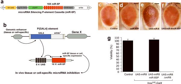
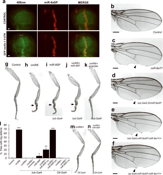
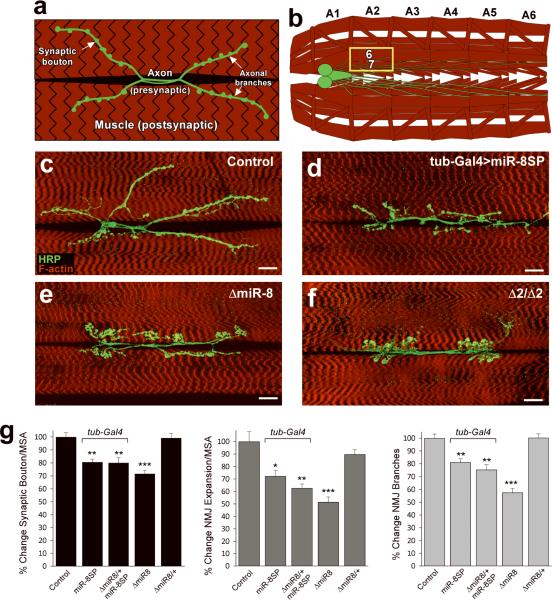
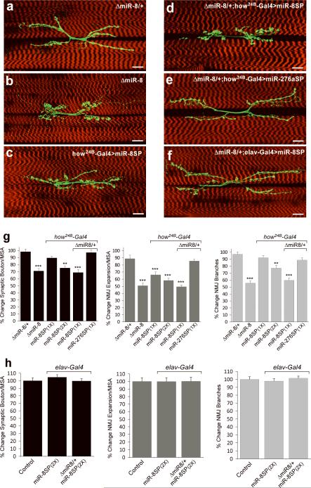
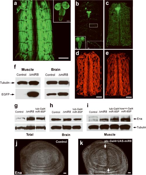
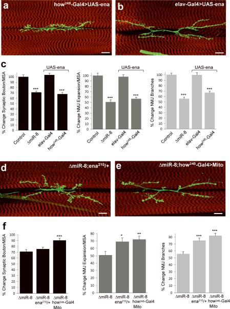
MicroRNAs are important regulators of gene expression, yet the functional outputs of most microRNA-target interactions remain elusive. Here we introduce the Drosophila melanogaster microRNA sponge (miR-SP) as a powerful transgenic technology to dissect the function of every microRNA with precise spatiotemporal resolution. miR-SPs can be used to characterize tissue-specific microRNA loss-of-function phenotypes, define the spatial regulation of their effectors and uncover interactions between microRNAs and other genes. Using themiR-SP system, we identified an essential role of the conserved microRNA miR-8, in neuromuscular junction formation. Tissue-specific silencing revealed that postsynaptic activity of miR-8 is important for normal neuromuscular junction morphogenesis. Given that miR-SPs rely on a bipartite modular expression system, they could be used to elucidate the endogenous function of microRNAs in any species in which conditional expression can be achieved.


Figures

FIGURE 1
FIGURE 1. Design and functionality of transgenic miR-SP elements
a, Ten microRNA binding sites (red) containing a mismatches at positions 9–12 were inserted downstream of EGFP (green) in a UAS-containing P-element vector. b, The resulting transgenic animals can be crossed to specific Gal4 lines. c–f, Expression of UAS-miR-8 results in a rough eye phenotype that can be rescued by co-expression of miR-8SP. Adult eye morphology in Control GMR-Gal4/+ (c), GMR-Gal4/miR-8SP;miR-8SP/+ (d), GMRGal4/+;UAS-miR-8/+ (e), GMR-Gal4/miR-8SP;UAS-miR-8/miR-8SP (f). Scale bars are 100 μm. g, Histogram illustrating percent viability in miR-8 null mutants and modified miR-8 genetic backgrounds (n > 50 for all genotypes; Error bars: ± SEM).
FIGURE 2
FIGURE 2. Effective tissue-specific silencing of endogenous microRNA activity by miR-SP
a, Wing imaginal discs from ptcGal4/miR-9aSP;miR-9aE39/+ (CONTROL) and tubulinEGFP nerfin-1 3'UTR, ptcGal4/miR-9aSP;miR-9aE39/+ (GFP nerfin-1 3'UTR,) imaged for GFP or mCherry. The cells along the anterior-posterior compartment boundary display increased GFP expression (brackets). Scale bars are 20 μm. b–f, MiR-9a silencing results in specific posterior wing margin defects. Cuticle preparations of adult wings from wild type (b), miR-9aJ22/miR-9aJ22 (c), miR-9aSP/+; miR-9aSP/tubulin-Gal4 (d), miR-9aSP/+; miR-9aJ22/tubulin-Gal4 (e), miR-9aSP/vg-Gal4; miR-9aJ22/+ (f). Scale bars are 100 μm. g–k, Inhibition of miR-8 activity by genomic knockdown or miR-SP silencing results in a deformed third leg pair. Cuticle preparations of adult third legs from wild type (g), ΔmiR-8miR-8 (h), miR-8SP/+; tubulin-Gal4/miR-8SP (i), miR-8SPmiR-8; tubulin-Gal4/miR-8SP (j), ΔmiR-8/+; tubulin-Gal4/Scramble-SP (k). l, Percentage of adult flies displaying malformed third leg phenotype in control and miR-8 modified genetic backgrounds (Student's t-test, *P=0.05; **P < 0.03; ***P < 10−4; Error bars: ± SEM). m, n, Cuticle preparation of adult third legs from Dll-Gal4miR-8 (m) and Dll-Gal4miR-8; miR-8SP/+ (n).
FIGURE 3
FIGURE 3. Discovery of novel microRNA functions using miR-SP elements
a,b, Schematic of the Drosophila third instar larval NMJ (a) and ventral body wall musculature (b). Abdominal segments are labeled A1–A6; anterior is left. The presynaptic compartment and CNS are shown in green and postsynaptic compartment in red; muscles 6 and 7 are highlighted in (a) and yellow box in (b). c–f, Immunofluorescence of larval NMJs from the indicated genotypes. All panels show confocal images of NMJ at muscles 6/7 in segment A2 of wandering third instar larvae stained for HRP (green, Horseradish Peroxidase) and F-actin (red). Scale bars are 20 μm. g, Quantification of synaptic bouton number, NMJ expansion and NMJ branch number as percentage of isogenized wild-type control. (*P<0.005; **P<0.001; ***P <10−7; by two-tailed Student's t-test; Error bars: mean ± SEM; n=20 for all genotypes and parameters). For NMJ analysis, statistical significance was determined with respect to the corresponding wild type or ΔmiR-8 heterozygous mutant background. Synaptic bouton numbers and NMJ expansion were normalized to muscle surface area (MSA). In all panels, “>” stands for driver-induced transgene expression.
FIGURE 4
FIGURE 4. MiR-SPs define microRNA activity with spatial specificity
a–f, Immunofluorescence of third instar larval NMJs from the indicated genotypes. Scale bars are 20 μm. g, h, Quantification of synaptic bouton number, NMJ expansion and NMJ branch number as percentage of isogenized wild-type control in a tissue-specific miR-8-modified genetic background in the post- (g) or pre-synaptic (h) compartments. ΔmiR-8/+ samples were statistically indistinguishable from wild-type NMJs and used as control reference for NMJ analysis of Δmir-8 heterozygous backgrounds. Post-synaptic miR-8SP expression results in a dose- dependent NMJ phenotype (g) similar to ΔmiR-8 null animals. In contrast, larvae expressing miR-8SP pan-neurally appeared statistically identical to wild-type controls (h) (Error bars are ± SEM; **P<0.0001; ***P<10−6 by two-tailed Student's t-test; n>17 for all genotypes and parameters)
FIGURE 5
FIGURE 5. MiR-SP elements can uncover tissue-specific microRNA function
a. EGFP under the control of a MiR-8 Gal4 driver is robustly expressed in the body wall musculature (a) and in the brain and ventral nerve cord (a, inset). b, c Confocal images of TubulinmiR-8-EGPF fluorescence in total dissected larvae (b, c), CNS (b solid inset) and motorneurons (b dashed inset) from wild-type controls compared to ΔmiR-8 mutants (c, e). Scale bars are 50 μm in (b) insets, and 500 μm in (d, e). f, Total miR-8-EGFP levels isolated from fresh muscle and brain extracts of control and ΔmiR-8 mutant animals, assessed by Western blotting using an anti-GFP antibody. g–i, Western blot analysis of Ena and tubulin expression in total dissected third instar larvae (g), CNS extracts (h) or muscle (i), in wild-type larvae (control), or larvae in which miR-8 was inhibited by ubiquitous expression of miR-8SP, or by homozygous miR-8 deletion (ΔmiR-8). Ten larvae were used to obtain the CNS homogenate in (h) and two larvae were used for muscle extracts in (i). j, k Third instar larval wing imaginal discs from wild type (j) and ptc-Gal4/UAS-miR-8 animals (k) immunostained with an Ena antibody. Cells expressing miR-8 along the anterior-posterior boundary, indicated by white arrows display reduced levels of Ena. Scale bars are 20 μm.
FIGURE 6
FIGURE 6. Genetic dissection of Ena function confirms miR-8-mediated postsynaptic regulation of NMJ morphogenesis
Analysis of NMJs at muscles 6/7 in larvae over-expressing UAS-ena under the muscle-specific how24B-Gal4 driver (a, c). The NMJs of larvae expressing UAS-ena pan-neurally under the control of the elav-Gal4 driver appear indistinguishable from wild-type controls (b, c). Reducing Ena levels by a heterozygous loss-of-function allele (d) or suppressing Ena activity postsynaptically by expressing UAS-FP4-mito (e) under the muscle-specific driver how24B-Gal4. Scale bars are 20 μm. Quantitatively, the synaptic bouton phenotype was rescued by 15 and 63%, NMJ expansion by 37 and 58%, and the reduction in branch number by 43 and 67% in the ena210 heterozygous and how24B-Gal4:UAS-FP4-mito backgrounds, respectively(f). (Error bars are ± SEM; for (c) ***P<10−6 relative to control; for (f) *P<0.005; **P<0.001; ***P<0.0001 relative to ΔmiR8 homozygous animals by two-tailed Student's t-test; n>17 for all genotypes and parameters).

Comment in

References

    1. Bushati N, Cohen SM. microRNA functions. Annu Rev Cell Dev Biol. 2007;23:175–205. - PubMed
    1. Eulalio A, Huntzinger E, Izaurralde E. Getting to the root of miRNA-mediated gene silencing. Cell. 2008;132:9–14. - PubMed
    1. Aboobaker AA, Tomancak P, Patel N, Rubin GM, Lai EC. Drosophila microRNAs exhibit diverse spatial expression patterns during embryonic development. Proc Natl Acad Sci U S A. 2005;102:18017–22. - PMC - PubMed
    1. Kawaji H, Hayashizaki Y. Exploration of small RNAs. PLoS Genet. 2008;4:e22. - PMC - PubMed
    1. Silver SJ, Hagen JW, Okamura K, Perrimon N, Lai EC. Functional screening identifies miR-315 as a potent activator of Wingless signaling. Proc Natl Acad Sci U S A. 2007;104:18151–6. - PMC - PubMed

Publication types

MeSH terms

LinkOut - more resources