. 2010 Feb 11;29(6):920-9.
doi: 10.1038/onc.2009.391. Epub 2009 Nov 16.

Cooperativity of the MUC1 oncoprotein and STAT1 pathway in poor prognosis human breast cancer

Affiliations

Cooperativity of the MUC1 oncoprotein and STAT1 pathway in poor prognosis human breast cancer

N Khodarev et al. Oncogene. .

Abstract

Signal transducer and activator of transcription 1 (STAT1) is activated in the inflammatory response to interferons. The MUC1 oncoprotein is overexpressed in human breast cancers. Analysis of genes differentially expressed in MUC1-transformed cells has identified a network linking MUC1 and STAT1 that is associated with cellular growth and inflammation. The results further show that the MUC1-C subunit associates with STAT1 in cells and the MUC1-C cytoplasmic domain binds directly to the STAT1 DNA-binding domain. The interaction between MUC1-C and STAT1 is inducible by IFNgamma in non-malignant epithelial cells and constitutive in breast cancer cells. Moreover, the MUC1-STAT1 interaction contributes to the activation of STAT1 target genes, including MUC1 itself. Analysis of two independent databases showed that MUC1 and STAT1 are coexpressed in about 15% of primary human breast tumors. Coexpression of MUC1 and the STAT1 pathway was found to be significantly associated with decreased recurrence-free and overall survival. These findings indicate that (i) MUC1 and STAT1 function in an auto-inductive loop, and (ii) activation of both MUC1 and the STAT1 pathway in breast tumors confers a poor prognosis for patients.


Conflict of interest statement

Dr. Kufe has an ownership interest in Genus Oncology and is a consultant to the company.The other authors disclosed no potential conflicts of interest.

Figures
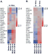
Figure 1
Figure 1. MUC1-CD activates transcription of STAT1 and STAT1-dependent genes
A. 3Y1/vector (clones A and B) and 3Y1/MUC1-CD (clones A and B) cells growing in vitro were analyzed for expression of the indicated genes in the IFNγ/STAT1 pathway. B. 3Y1/MUC1-CD cells growing in vitro and as tumors in nude mice were analyzed for expression of the indicated IFNγ/STAT1 target genes. Values are in log2 scale and represent expression relative to the average value of the gene across samples.
Figure 2
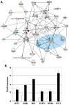
Figure 2. Functional gene network analysis links MUC1 and STAT1
A. Genes differentially expressed in 3Y1/MUC1-CD cells growing in vivo as compared to in vitro were analyzed using Ingenuity Pathway Analysis to identify functional networks. The gene network containing MUC1 and STAT1 as shown is associated with genes involved in cellular growth and inflammation. Red signifies up-regulation. Green signifies down-regulation. B. 3Y1/vector (open bars) and 3Y1/MUC1-CD (solid bars) were left untreated and treated with IFNγ for 24 h. Total RNA was analyzed by quantitative RT-PCR for expression of the indicated genes. The results are expressed as the fold-induction (mean±SD) for IFNγ-treated as compared to untreated cells. The differences between values obtained for the 3Y1/vector and 3Y1/MUC1-CD cells were significant at p values ranging from 0.035 to 0.00078.
Figure 3
Figure 3. MUC1-C interacts directly with STAT1
A. Lysates from 3Y1/MUC1-CD cells were immunoprecipitated with anti-MUC1-C or a control IgG. The precipitates were immunoblotted with the indicated antibodies. B. Amino acid sequence of MUC1-CD and MUC1-CD (mSRM) (upper panel). GST, GST-MUC1-CD, GST-MUC1-CD (1–45) and GST-MUC1-CD (46–72) bound to glutathione beads were incubated with purified recombinant STAT1 (lower left). GST, GST-MUC1-CD and GST-MUC1-CD (mSRM) bound to gluthatione beads were incubated with recombinant STAT1 (lower right). The adsorbates were immunoblotted with anti-STAT1. Input of the GST and GST-MUC1-CD proteins was assessed by Coomassie blue staining. C. Structure of STAT1 (upper panel). GST, GST-STAT1 (full length; amino acids 1–750), GST-STAT1 (N-terminal; amino acids 1–324), GST-STAT1 (DBD; amino acids 295–491) and GST-STAT1 (C-terminal; amino acids 464–750) bound to glutathione beads were incubated with purified MUC1-CD. Adsorbates were immunoblotted with anti-MUC1-C. Input of GST and GST-STAT1 fusion proteins was assessed by Coomassie blue staining.
Figure 4
Figure 4. Induction of MUC1 by IFNγ in MCF-10A cells is conferred by both STAT1 and MUC1
A. MCF-10A cells were treated with IFNγ for the indicated times. Lysates were immunoblotted with the indicated antibodies (left). MUC1 and, as a control, β-actin mRNA levels were determined by RT-PCR (right). B. MCF-10A cells left untreated or treated with IFNγ for 24 h were immunoprecitated with anti-STAT1. The precipitates were immunoblotted with the indicated antibodies. C. MCF-10A cells were transfected with control or STAT1 siRNA pools for 24 h and then left untreated or stimulated with IFNγ for 24. Lysates were immunoblotted with the indicated antibodies. D. MCF-10A cells were left untreated and treated with 5 μM GO-201 or CP-1 for 3 days and then stimulated with IFNγ for 24 h. Whole cell lysates were immunoblotted with the indicated antibodies. NS: non-specific band. E. MCF-10A cells were transfected with control or MUC1siRNA pools for 72 h. The transfected cells were left untreated or stimulated with IFNγ for 24 h. Total cellular RNA was analyzed for IFITM1 expression by real-time RT-PCR. The results are expressed as the fold activation (mean±SD from three experiments) compared with that obtained with cells transfected with the control siRNA and left untreated (assigned a value of 1).
Figure 5
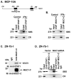
Figure 5. MUC1-C associates with the STAT1 transcription complex
A. Soluble chromatin from MCF-10A cells left untreated or treated with IFNγ for 24 h was immunoprecipitated with anti-STAT1. The final DNA extractions were amplified by PCR with pairs of primers that cover the STAT binding site (SBS; −689 to −414) and the control region (CR; +4524 to +4745) in the MUC1 promoter. B. Soluble chromatin from MCF-10A cells left untreated or treated with IFNγ for 24 h was immunoprecipitated with anti-MUC1-C and analyzed for SBS and CR sequences. C. Lysates from ZR-75-1 cells were immunoprecipitated with anti-MUC1-C or a control IgG. The precipitates were immunoblotted with the indicated antibodies. D. Soluble chromatin from ZR-75-1/vector and ZR-75-1/MUC1siRNA cells was precipitated anti-STAT1 and analyzed for MUC1 promoter SBS and CR sequences. In re-ChIP analysis, the anti-STAT1 precipitates were released, reimmunoprecipitated with anti-MUC1-C and then analyzed for MUC1 promoter sequences.
Figure 6
Figure 6. Coexpression of MUC1 and the STAT1 pathway is associated with reduced recurrence-free survival
A. An expressional database derived from 327 breast tumors was analyzed for expression of MUC1 (MUC1+) and STAT1 (STAT1+). Coexpression was detectable in 45 tumors (13.8%). Hierarchical clustering for expression of the STAT1 pathway (24 genes) was performed for the entire database. Relative expression values are in log2 scale. Activation of the STAT1 pathway (STAT1P+) was significantly associated with MUC1 expression (49 tumors; Fisher’s exact test, P=0.0003). B. Kaplan-Meier curves for recurrence-free survival were determined for patients with MUC1+/STAT1P+ tumors (n=49; red curve) compared to those patients without coexpression (n=257; blue curve). C. Kaplan-Meier curves were determined for recurrence-free survival of patients with grade 2/3 tumors that are MUC1+/STAT1P+ (n=32; red curve) compared to those patients with grade 2/3 tumors without coexpression (n=169; blue curve).
Figure 7
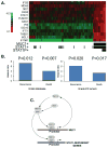
Figure 7. STAT1 pathway activation linked to MUC1 expression is associated with increased risk of death
A. An additional expressional database derived from 155 breast tumors was analyzed for expression of MUC1 (MUC1+) and STAT1 (STAT1+). Coexpression was detectable in 25 tumors (16.1%). Hierarchical clustering for expression of the STAT1 pathway (24 genes) was performed for the MUC1+ tumors. Relative expression values are in log2 scale. Activation of the STAT1 pathway (STAT1P+) was significantly associated with MUC1 expression (P<0.0001). Genes having significantly increased expression in MUC1+ tumors are shown. B. Hazard ratios for recurrence-free and overall survival were determined for patients with MUC1+/STAT1P+ tumors (n=8) compared to those patients without coexpression (n=147) (left panel). Hazard ratios for recurrence-free and overall survival were determined for patients with grade 2/3 tumors that are MUC1+/STAT1P+ (n=8) compared to those patients with grade 2/3 tumors without coexpression (n=126) (right panel). C. Proposed schema for a MUC1-STAT1 autoinductive loop.

References

    1. Ahmad R, Raina D, Joshi MD, Kawano T, Kharbanda S, Kufe D. MUC1-C oncoprotein functions as a direct activator of the NF-kappaB p65 transcription factor. Cancer Res. 2009;69:7013–7021. - PMC - PubMed
    1. Ahmad R, Raina D, Trivedi V, Ren J, Rajabi H, Kharbanda S, et al. MUC1 oncoprotein activates the I| B kinase β complex and constitutive NF-| B signaling. Nat Cell Biol. 2007;9:1419–1427. - PMC - PubMed
    1. Chanrion M, Negre V, Fontaine H, Salvetat N, Bibeau F, Mac Grogan G, et al. A gene expression signature that can predict the recurrence of tamoxifen-treated primary breast cancer. Clin Cancer Res. 2008;14:1744–1752. - PMC - PubMed
    1. Darnell JE, Jr, Kerr IM, Stark GR. Jak-STAT pathways and transcriptional activation in response to IFNs and other extracellular signaling proteins. Science. 1994;264:1415–1421. - PubMed
    1. Deblandre GA, Marinx OP, Evans SS, Majjaj S, Leo O, Caput D, et al. Expression cloning of an interferon-inducible 17-kDa membrane protein implicated in the control of cell growth. 1995;270:23860–23866. - PubMed

Publication types

MeSH terms