. 2010 Feb 11;29(6):811-21.
doi: 10.1038/onc.2009.389. Epub 2009 Nov 16.

Runx2 association with progression of prostate cancer in patients: mechanisms mediating bone osteolysis and osteoblastic metastatic lesions

Affiliations

Runx2 association with progression of prostate cancer in patients: mechanisms mediating bone osteolysis and osteoblastic metastatic lesions

J Akech et al. Oncogene. .

Abstract

Runx2, a bone-specific transcriptional regulator, is abnormally expressed in highly metastatic prostate cancer cells. Here, we identified the functional activities of Runx2 in facilitating tumor growth and osteolysis. Our studies show that negligible Runx2 is found in normal prostate epithelial and non-metastatic LNCaP prostate cancer cells. In the intra-tibial metastasis model, high Runx2 levels are associated with development of large tumors, increased expression of metastasis-related genes (MMP9, MMP13, VEGF, Osteopontin) and secreted bone-resorbing factors (PTHrP, IL8) promoting osteolytic disease. Runx2 siRNA treatment of PC3 cells decreased cell migration and invasion through Matrigel in vitro, and in vivo shRunx2 expression in PC3 cells blocked their ability to survive in the bone microenvironment. Mechanisms of Runx2 function were identified in co-culture studies showing that PC3 cells promote osteoclastogenesis and inhibit osteoblast activity. The clinical significance of these findings is supported by human tissue microarray studies of prostate tumors at stages of cancer progression, in which Runx2 is expressed in both adenocarcinomas and metastatic tumors. Together these findings indicate that Runx2 is a key regulator of events associated with prostate cancer metastatic bone disease.


Figures

Figure 1
Figure 1. Runx2 expression in prostate cancer cell lines regulates metastatic properties
(A) Runx2 mRNA levels in PC3, LNCaP, C4-2B and non-tumorigenic RWPE cells were analyzed by qRT-PCR (upper panel, see Methods). Protein levels by western blot analysis (lower panel). (B) PC3 sublines (PC3-H, PC3-M, PC3-L) were analyzed for Runx2 mRNA and protein in whole cell lysates and nuclear extracts. (C) Western blot shows 80% knockdown of endogenous Runx2 in PC3-H cells; CDK1 is used as internal control. (D) Invasion through matrigel is reduced and adhesion increased in Runx2 siRNA treated PC3-H cells. (E) Expression of Runx2 and target genes (OC, VEGF and MMP9) by qRT-PCR following knockdown (48hr). Values are mean ± SD of n=3 samples analyzed in triplicate. (F) Reduced expression of survivin by Runx2 siRNA. E, F panels are representative of triplicate assays of n=4 experiments. Error bars are mean ± SD.
Figure 2
Figure 2. Runx2 expression in PC3 sublines promotes osteolytic bone disease in vivo and osteoclast activation in vitro
(A) Mouse tibiae at 4 and 6 weeks expressing different Runx2 levels (PC3-H, PC3-M, PC3-L) as indicated. Aggressive osteolysis in tumors from PC3-H cells (at 4 weeks); near complete erosion through cortical bone also occurs in PC3-M. PC3-L induced tumors have minimal lysis at 4 weeks and increased radiopaque areas at 6 weeks which fill in the eroded areas and represent osteoblastic lesions. Quantification of extent of osteolysis marked areas in PC3-H and PC3-M is shown (B) PC3 cells induce osteolytic lesions shown by TRAP staining of the bone resorbing cells (arrow shows osteoclasts). PC3-L tumors induced woven bone (WB) formation, shown here at the tumor-bone interface. Immunohistochemistry of the tumor area for Runx2 expression in PC3 tumors. The human specific marker cytokeratin (10x) and Runx2 is shown (40x). Insets show IgG antibody control. (C) Expression of Runx2 by qRT-PCR showing mRNA levels in PC3 sublines post-implantation. Data shown are mean ± SD of samples analyzed in triplicates. (D) PTHrP and IL8 expression in PC3 sublines measured by qRT-PCR.
Figure 3
Figure 3
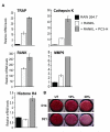
(A) Gene expression of markers for osteoclast differentiation detected by qRT-PCR after co-culturing PC3-H cells with RAW 264.7 cells. (B) Histochemical staining of MC3T3 cells for alkaline phosphatase shows inhibited osteoblast differentiation when medium from PC3-H cells (10% or 20%) was added to cultured osteoblasts and analyzed after 14 and 21 days.
Figure 4
Figure 4. Forced expression of Runx2 leads to activation of metastatic markers
(A) Expression of MMP9, MMP13, MMP2, VEGF, OP and OC in PC3 sublines by qRT-PCR. GAPDH was used for internal control for qRT-PCR. Data shown are mean ± SD of samples analyzed in triplicates (B) Expression levels of metastasis related genes in PC3-M, PC3-L and LNCaP cells after viral transduction are shown and were normalized to GAPDH. Runx2 regulation of MMP2, MMP9, MMP13, OC and OP is shown. Data from one representative of n=4 experiments is shown. Values are mean ± SD.
Figure 5
Figure 5. Runx2 knockdown reduces metastatic bone disease
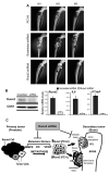
(A) Radiographs of tibiae 3 weeks post-injection of tumor cells show osteolytic lesions caused by parental PC3-H cells (top panel) and slightly more aggressive lesions by PC3-H cells infected with scrambled shRNA (center), but absence of tumor activity (M1, M3) or evidence of onset of lysis (M2) in the Runx2 shRNA group (lower panels, arrows point to onset of lysis). (B) Western blot showing Runx2 protein levels of whole cell extracts from PC3-H infected with scrambled or Runx2 shRNA (left). qRT-PCR showing knockdown of Runx2, IL8 and PTHrP in tumors from implanted PC3-H cells infected with scrambled or Runx2 shRNA (right). (C) Runx2 mediated mechanisms of metastasis and formation of osteolytic and osteoblastic bone lesions. Prostate cancer progression from primary prostate tumor to metastatic cancer occurs through Runx2 regulation of metastasis related genes (e.g. MMP9, OC, VEGF, see Figs 1 and 4). At the onset of bone metastasis, tumor cells become heterogeneous, expressing different levels of Runx2 [high Runx2 (PC3-H) and low Runx2 (PC3-L)]. Within the tumor-bone interface, PC3-H cells release factors (PTHrP, IL8, see Fig 2D) that activate osteoclasts (OCL) resulting in bone resorption and osteolytic lesions. PC3-L cells form osteoblastic lesions induce new bone formation potentiated by osteoblasts (OB) via several possible pathways [reviewed in (Keller and Brown, 2004), and in discussion]. Runx2 knockdown (siRNA; shRNA in vivo) blocks the expression of metastasis promoting genes and reduces the formation of osteolytic lesions.

References

    1. Altieri DC. New wirings in the survivin networks. Oncogene. 2008;27:6276–6284. - PMC - PubMed
    1. Araki S, Omori Y, Lyn D, Singh RK, Meinbach DM, Sandman Y, et al. Interleukin-8 is a molecular determinant of androgen independence and progression in prostate cancer. Cancer Res. 2007;67:6854–6862. - PubMed
    1. Armstrong AP, Miller RE, Jones JC, Zhang J, Keller ET, Dougall WC. RANKL acts directly on RANK-expressing prostate tumor cells and mediates migration and expression of tumor metastasis genes. Prostate. 2008;68:92–104. - PubMed
    1. Barnes GL, Hebert KE, Kamal M, Javed A, Einhorn TA, Lian JB, et al. Fidelity of Runx2 activity in breast cancer cells is required for the generation of metastases associated osteolytic disease. Cancer Res. 2004;64:4506–4513. - PubMed
    1. Barnes GL, Javed A, Waller SM, Kamal MH, Hebert KE, Hassan MQ, et al. Osteoblast-related transcription factors Runx2 (Cbfa1/AML3) and MSX2 mediate the expression of bone sialoprotein in human metastatic breast cancer cells. Cancer Res. 2003;63:2631–2637. - PubMed

Publication types

MeSH terms

Substances