. 2010 Feb 11;29(6):930-6.
doi: 10.1038/onc.2009.368. Epub 2009 Nov 16.

Enhanced mRNA cap methylation increases cyclin D1 expression and promotes cell transformation

Affiliations

Enhanced mRNA cap methylation increases cyclin D1 expression and promotes cell transformation

V H Cowling. Oncogene. .

Abstract

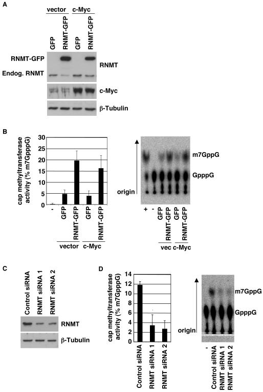
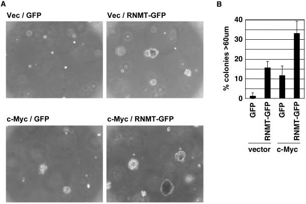
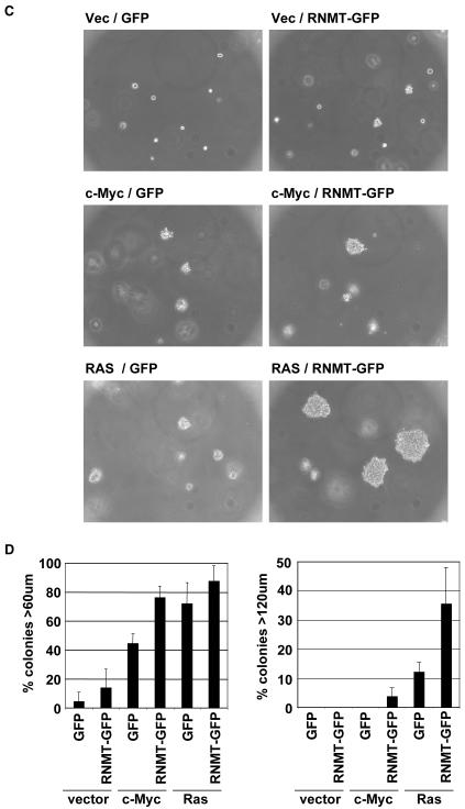
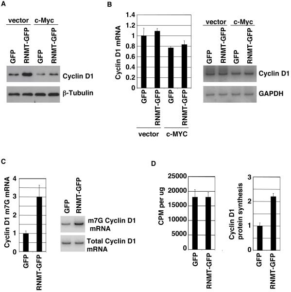
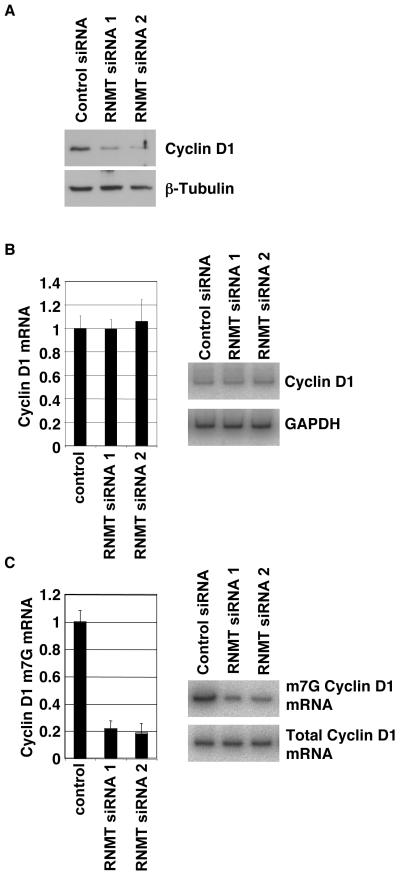
Cap-dependent mRNA translation requires the methylation of the mRNA guanosine cap by RNA guanine-7-methyltransferase (RNMT). mRNA cap methylation was recently described to be rate-limiting for a subset of mRNAs, and to be enhanced by expression of c-Myc and E2F1, although the biological significance of this finding was not investigated. Here, it is reported that increased RNMT expression enhances cellular mRNA cap methyltransferase activity, promotes mammary epithelial cell transformation and cooperates with H-RasV12 or c-Myc to promote fibroblast cell transformation. Cyclin D1 is a prominent oncogene in epithelial tumours. A significant fraction of Cyclin D1 mRNA was found to be unmethylated on the mRNA cap and thus dormant in mammary epithelial cells. Cyclin D1 expression was increased by enhanced mRNA cap methylation. In summary, this report shows that mRNA cap methylation is rate-limiting for expression of an oncogene and cell transformation.


Figures

Figure 1
Figure 1. Increased RNMT (RNA guanine-7-methyltransferase) expression increases cellular mRNA cap methyltransferase activity
a) Whole cell extracts from the IMEC (Tert-immortalised human mammary epithelial cell) lines indicated were analysed by Western Blot for expression of RNMT, RNMT-GFP, c-MYC and β-Tubulin. The migration of endogenous RNMT and RNMT-GFP is indicated. Cells were lysed in Triton lysis buffer (10 mM Tris, pH 7.05, 50 mM NaCl, 30 mM Na4 pyrophosphate, 50 mM NaF, 5 μM ZnCl2, 10% glycerol, 0.5% Triton X-100, 10μM Leupeptin, 1μM Pepstatin and 0.1mg/ml Aprotinin). Polyclonal anti-RNMT antibodies were raised against full length recombinant GST-RNMT in sheep and affinity-purified and other antibodies were purchased from Santa Cruz Biotechnologies (Santa Cruz, CA, USA). b) The same extracts were analysed for their cap methyltransferase activity. Assay was adapted from (Pillutla et al., 1998). A 55 base strand of in vitro transcribed RNA was capped using Vaccina Virus capping enzyme and 32P alpha-GTP according to manufacturers instructions (Epicentre Biotechnologies, Madison, WI, USA). 10-40ng G32P-RNA was incubated with 2μg cell extract in 10μl 50mM Tris pH 8, 6mM KCl, 1.25mM MgCl2, 100nM S-adenosyl methionine at 37°C for 10mins. RNA was purified, precipitated and resuspended in 4μl 50mM NaAcetate and 0.25units P1 nuclease for 30mins at 37°C. Cap (GpppG) and methyl-cap (m7GpppG) were resolved in 0.4M Ammonium Sulphate thin layer chromatography using PEI cellulose plates. Standards were visualised by UV light to establish correct migration. Labelled spots were visualised and quantified by autoradiography, and percentage conversion of GpppG to m7GppG was calculated. Average results for three independent experiments are shown and error bars indicate the standard deviation. A representative assay is also shown including a negative control (-), in which no extract was added and a positive control (+), in which recombinant RNMT was added. c) IMEC cells were transfected with two independent siRNAs directed against RNMT or Cyclophilin B (negative control), according to manufacturer’s instructions (Dharmacon Inc, Lafayette, CO, USA). After 24hrs, cell extracts were prepared and analysed for their cap methyltransferase activity, as above, except 4μg of cell extract was used.
Figure 2
Figure 2. RNMT promotes cell transformation
a,b) IMEC cells and c,d) Rat1A fibroblasts expressing the proteins indicated were used in the soft agar transformation assay, performed according to (Cowling et al., 2006) with the following modifications. 2.5 × 10⁴ IMECs/ml were plated in 0.3% noble agar/IMEC medium supplemented with 5% FBS (Invitrogen). Cells were fed with 250μl IMEC medium/5% FBS every 2nd day, starting with the day of plating. Rat1A cells were plated in DMEM/10% FCS and fed every 4th day, starting with the day of plating. a, c) Micrographs of colonies at 14 and 7 days, respectively. b, d) After 7 days the size of colonies was measured for 100 plated cells using a graticle. Chart depicts the percentage of cells developing into colonies of the size indicated at 7 days. Average results for three independent experiments are shown and error bars indicate the standard deviation.
Figure 3
Figure 3. RNMT promotes Cyclin D1 mRNA cap methylation and translation
All experiments were performed in the IMEC lines indicated. a) Expression of Cyclin D1 and β-Tubulin was analysed by Western Blot. b) Cyclin D1 mRNA levels were determined by RT-PCR performed using Superscript One Step RT-PCR System (Invitrogen, Carlsbad, CA, USA). Reactions were labelled using 32P labelled-primers, resolved by gel electrophoresis and quantified by phosphoimager. A representative gel is shown. Reactions were determined to be in the linear range by performing a titration of input mRNA. Reactions were normalised to rRNA levels since the cellular rRNA content was unchanged in response to expression of RNMT. c) Cyclin D1 m7G mRNA levels were determined by anti-m7G antibody immunoprecipitation followed by RT-PCR, in the cell lines indicated, performed according to (Cole and Cowling, 2009). A representative gel is shown. d) The IMEC lines were labelled with 35S cysteine and methionine and incorporation per μg protein extract and into Cyclin D1 was determined. 10cm plates of IMECs were incubated in 4ml regular growth medium minus cysteine and methionine containing 10μM MG132 for 30mins at 37°C. 220μCi Express Protein Labelling Mix containing 35S cysteine and methionine (Perkin Elmer) was added for 15mins. Cells were lysed, normalised for protein content and 35S-cysteine and methionine incorporation into TCA-precipitated protein was detected using a scintillation counter. Immunoprecipitations performed on half of each extract using 2.5μl polyclonal anti-Cyclin D1 or control polyclonal antibodies were resolved by gel electrophoresis. Label incorporation into Cyclin D1 was detected by phosphoimager and expressed as values relative to input. For all charts, average results for three independent experiments are shown and error bars indicate the standard deviation.
Figure 4
Figure 4. RNMT is necessary for Cyclin D1 mRNA cap methylation and translation
IMEC cells were transfected with two independent siRNAs directed against RNMT or Cyclophilin B (negative control), as in figure 1. After 24hrs, cells were lysed and protein and RNA was extracted. a) Whole cell extracts were analysed by Western Blot for expression of RNMT, Cyclin D1 and β-Tubulin. RNA was analysed for relative expression level of b) total Cyclin D1 mRNA and c) Cyclin D1 m7G mRNA. Representative gels are shown. For all charts, average results for three independent experiments are shown and error bars indicate the standard deviation.

References

    1. Bentley DL. Rules of engagement: co-transcriptional recruitment of pre-mRNA processing factors. Curr Opin Cell Biol. 2005;17:251–6. - PubMed
    1. Chu C, Shatkin AJ. Apoptosis and autophagy induction in mammalian cells by small interfering RNA knockdown of mRNA capping enzymes. Mol Cell Biol. 2008;28:5829–36. - PMC - PubMed
    1. Cole MD, Cowling VH. Transcription-independent functions of MYC: regulation of translation and DNA replication. Nat Rev Mol Cell Biol. 2008;9:810–5. - PMC - PubMed
    1. Cole MD, Cowling VH. Specific regulation of mRNA cap methylation by the c-Myc and E2F1 transcription factors. Oncogene. 2009 - PMC - PubMed
    1. Cowling VH, Chandriani S, Whitfield ML, Cole MD. A conserved Myc protein domain, MBIV, regulates DNA binding, apoptosis, transformation, and G2 arrest. Mol Cell Biol. 2006;26:4226–39. - PMC - PubMed

Publication types