. 2009 Nov 12;4(11):e7804.
doi: 10.1371/journal.pone.0007804.

Nitric oxide releasing nanoparticles are therapeutic for Staphylococcus aureus abscesses in a murine model of infection

Affiliations

Nitric oxide releasing nanoparticles are therapeutic for Staphylococcus aureus abscesses in a murine model of infection

George Han et al. PLoS One. .

Abstract

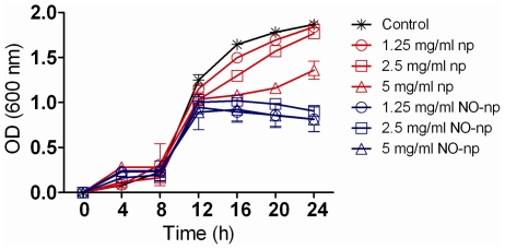
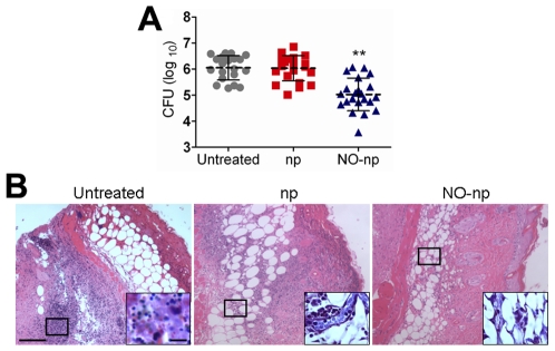
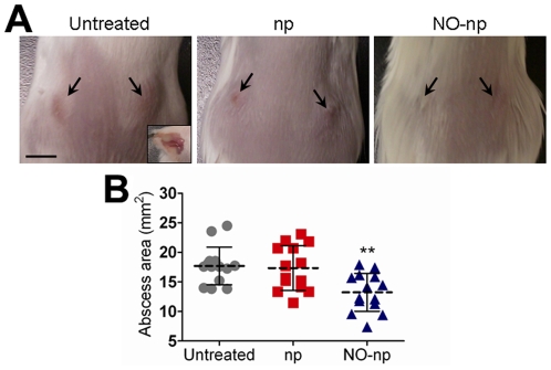
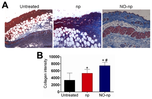
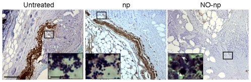
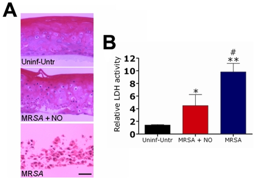
Staphylococcus aureus (SA) is a leading cause of a diverse spectrum of bacterial diseases, including abscesses. Nitric oxide (NO) is a critical component of the natural host defense against pathogens such as SA, but its therapeutic applications have been limited by a lack of effective delivery options. We tested the efficacy of a NO-releasing nanoparticle system (NO-np) in methicillin-resistant SA (MRSA) abscesses in mice. The results show that the NO-np exert antimicrobial activity against MRSA in vitro and in abscesses. Topical or intradermal NO-np treatment of abscesses reduces the involved area and bacterial load while improving skin architecture. Notably, we evaluated pro- and anti-inflammatory cytokines that are involved in immunomodulation and wound healing, revealing that NO-np lead to a reduction in angiogenesis preventing bacterial dissemination from abscesses. These data suggest that NO-np may be useful therapeutics for microbial abscesses.


Conflict of interest statement

Competing Interests: The authors have declared that no competing interests exist.

Figures

Figure 1
Figure 1. NO-np inhibits MRSA growth in vitro.
The effect of NO in MRSA growth kinetics after 24 h co-culture was determined using Bioscreen C analysis. MRSA was grown in the absence or presence of nanoparticles with NO (NO-np) or without NO (np). Each point represents the average of four measurements and error bars denote standard deviations.
Figure 2
Figure 2. NO-np killed MRSA in subcutaneous abscesses.
(A) Abscess bacterial burden (CFU; colony forming units) in mice infected subcutaneously with 107 MRSA and treated with NO-np is significantly lower than untreated or np-treated mice (n = 20 abscesses per group). Each point represents an abscess. Dashed lines are the averages of the results and error bars denote standard deviations. Asterisks denote P value significance (** P<0.001) calculated by analysis of variance and adjusted by use of the Bonferroni correction. (B) Histological analysis of Balb/c mice untreated MRSA-infected, np-treated MRSA-infected, and MRSA-infected treated with NO, day 4. Mice were infected with 107 MRSA. Representative H&E-stained sections of the skin lesions are shown with the Insets showing Gram staining of MRSA. Scale bars: 25 µm.
Figure 3
Figure 3. NO-np decreases subcutaneous abscess area in mice.
(A) Abscesses of Balb/c mice untreated MRSA-infected, np-treated MRSA-infected, and MRSA-infected treated with NO-np, day 4. Arrows denote abscesses. Inset shows a representative purulent abscess 4 days after MRSA infection. Scale bar: 5 mm. (B) Abscess area analysis of Balb/c mice subcutaneous lesions. Abscesses were infected with MRSA and untreated or treated in the absence or presence of NO. Each point represents an abscess. Dashed lines are the averages of the results for fourteen measurements, and error bars denote standard deviations. Asterisks denote P value significance (** P<0.001) calculated by analysis of variance and adjusted by use of the Bonferroni correction.
Figure 4
Figure 4. NO-np prevents MRSA collagen degradation in subcutaneous abscesses.
(A) Histological analysis of Balb/c mice untreated MRSA-infected, np-treated MRSA-infected, and MRSA-infected treated with NO-np, day 4. Mice were infected with 107 bacterial cells. The blue stain indicates collagen. Scale bar: 25 µm. (B) Quantitative measurement of collagen intensity in 20 representative fields of the same size for untreated MRSA-infected, np-treated MRSA-infected, and MRSA-infected treated with NO-np. Bars are the averages of the results, and error bars denote standard deviations. Asterisks denote P value significance (*, P<0.05 in comparing MRSA-infected np and untreated control groups; **, P<0.001 in comparing MRSA-infected NO-np and untreated control groups; #, P<0.01 in comparing MRSA-infected np and NO-np groups) calculated by analysis of variance and adjusted by use of the Bonferroni correction.
Figure 5
Figure 5. NO-np decreases vascularization in the setting of MRSA abscesses.
Histological analysis of untreated, np-treated and NO-np treated MRSA-infected Balb/c mice, day 4. Mice were infected with 107 MRSA. The brown staining indicates vascularization. Representative CD34-immunostained sections of the skin lesions are shown with the Insets representing Gram of MRSA. White arrows denote Gram positive cocci. Scale bars: 25 µm.
Figure 6
Figure 6. NO reduce cellular damage of reconstituted epidermal tissue infected with MRSA.
(A) NO-np attenuate cellular damage of reconstituted epidermal tissue infected with MRSA. Reconstituted human epidermis control group had no bacteria or nanoparticles, reconstituted human epidermis infected with SA, and reconstituted human epidermis infected with SA and treated with NO-np. Scale bar, 10 µm. (B) Relative LDH activity measured in the tissue culture supernatant by reconstituted human epidermal tissue after 24 h co-culture with SA in the absence or presence of NO-np. Bars are the averages of the results for three measurements, and error bars denote standard deviations. Asterisks denote P value significance (*, P<0.05 in comparing the MRSA+NO-np group with the control group; **, P<0.001 in comparing the MRSA group with control group; #, P<0.01 in comparing the MRSA group with MRSA+NO-np group) calculated by analysis of variance and adjusted by use of the Bonferroni correction.

Similar articles

Cited by

References

    1. McCaig LF, McDonald LC, Mandal S, Jernigan DB. Staphylococcus aureus-associated skin and soft tissue infections in ambulatory care. Emerg Infect Dis. 2006;12:1715–1723. - PMC - PubMed
    1. Noskin GA, Rubin RJ, Schentag JJ, Kluytmans J, Hedblom EC, et al. National trends in Staphylococcus aureus infection rates: impact on economic burden and mortality over a 6-year period (1998-2003). Clin Infect Dis. 2007;45:1132–1140. - PubMed
    1. Moran GJ, Krishnadasan A, Gorwitz RJ, Fosheim GE, McDougal LK, et al. Methicillin-resistant S. aureus infections among patients in the emergency department. N Engl J Med. 2006;355:666–674. - PubMed
    1. Hiramatsu K, Hanaki H, Ino T, Yabuta K, Oguri T, et al. Methicillin-resistant Staphylococcus aureus clinical strain with reduced vancomycin susceptibility. J Antimicrob Chemother. 1997;40:135–136. - PubMed
    1. Klevens RM, Morrison MA, Nadle J, Petit S, Gershman K, et al. Invasive methicillin-resistant Staphylococcus aureus infections in the United States. JAMA. 2007;298:1763–1771. - PubMed

Publication types