. 2009 Nov 13;4(11):e7825.
doi: 10.1371/journal.pone.0007825.

Augmentation of reverse transcription by integrase through an interaction with host factor, SIP1/Gemin2 Is critical for HIV-1 infection

Affiliations

Augmentation of reverse transcription by integrase through an interaction with host factor, SIP1/Gemin2 Is critical for HIV-1 infection

Hironori Nishitsuji et al. PLoS One. .

Abstract

There has been accumulating evidence for the involvement of retroviral integrase (IN) in the reverse transcription of viral RNA. We previously identified a host factor, survival motor neuron-interacting protein 1 (SIP1/Gemin2) that binds to human immunodeficiency virus type 1 (HIV-1) IN and supports HIV-1 infection apparently at reverse transcription step. Here, we demonstrated that HIV-1 IN together with SIP1 augments reverse transcriptase (RT) activity by enhancing the assembly of RT on viral RNA in vitro. Synthetic peptides corresponding to the binding motifs within IN that inhibited the IN-SIP1 interaction abrogated reverse transcription in vitro and in vivo. Furthermore, knockdown of SIP1 reduced intracellular stability and multimer formation of IN through proteasome-mediated degradation machinery. Taken together, SIP1 appears to stabilize functional multimer forms of IN, thereby promoting the assembly of IN and RT on viral RNA to allow efficient reverse transcription, which is a prerequisite for efficient HIV-1 infection.


Conflict of interest statement

Competing Interests: The authors have declared that no competing interests exist.

Figures

Figure 1
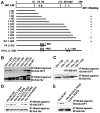
Figure 1. Intracellular interaction between SIP1 and IN mutants resulting in a reverse transcription defective phenotype.
(A) Schematic diagram of HIV-1 integrase. Numbers refer to amino-acid residues of IN. The locations of the critical amino acids for reverse transcription in vivo (Tyr15, Lys 186, Arg187 and Lys188, and LeuLeu241,242) and the active site residues Asp116 are indicated. The region of each truncated form of IN is shown as a solid line. (B, C) Wild type (WT) or IN mutant expression plasmids with a V5-tag were transfected into 293T cells. For transfection, 1.0 µg of each plasmid for WT-V5, D116G, N-ter, Cen, C-ter, N-Cen or Cen-C, and 2.5 µg for Y15A, K186Q, delta-KRK or LL241,242AA was used, respectively. At 48 h after transfection, cells were harvested and lysed with RSB-100 containing 1.0% NP-40. Cell lysate was then subjected to immunoprecipitation using an anti-V5 antibody, followed by Western blot analysis with an anti-SIP1 antibody. H and L denote heavy and light chains of immunoglobulin, respectively.
Figure 2
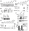
Figure 2. Residues of HIV-1 IN that are critical for specific interactions with recombinant SIP1 in vitro.
(A) A schematic of the full length (open bar, top) of HIV-1 IN and its deletion forms (solid line) are shown. Point mutations of the residues of IN (Val75, Ala76, Leu241 and Leu242) that are critical for interaction with SIP1, are indicated with their location numbers. (B, C) For pull-down experiments, 10 µg of recombinant SIP1 was incubated with 10 µg of each His-tagged IN (His-IN), followed by purification through a nickel agarose column. The level of SIP1 bound to each IN-nickel agarose complex was determined by Western blot analysis using an anti-SIP1 antibody. (D) Series of amino acid substitutions targeting the residues from 71 to 80 of the His-tagged IN (1–90) protein were generated. Interaction of each mutant protein with SIP1 was determined as described above in (B). (E) Full-length His-tagged IN mutant proteins carrying the VA74,75AG or LL241,242AA mutations were generated. Interaction of each mutant protein with SIP1 was determined as described above in (B).
Figure 3
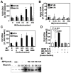
Figure 3. Characterization of intracellular IN proteins.
(A) 293T cells were transfected with indicated plasmid in presence of 10 µM MG-132 (+) or DMSO(−). Total cell lysates (10 µg) were analyzed by Western blotting analysis. (B, C) 293T cells were transfected twice with 100 pmol of siControl or siSIP1 and then with lacZ-V5 or an IN-V5 expression plasmid with MG-132 or DMSO. 48 h later, cells were lysed with CSK buffer containing 0.5% NP-40 followed by Western blotting analysis. (D, E) 293T cells transfected each IN-V5 expression vector were suspended with CSK buffer containing 0.5% NP-40. Cell lysates were incubated in either the absence or presence of 0.2 mM DTSSP at room temperature for 20 min and analyzed by Western blotting using an anti-V5 antibody. (F) Cell lysates in (B) were treated with 0.2 mM of DTSSP and then analyzed by Western blotting. The relative ratio of monomers, dimers, and tetramers after treatment with 0.2 mM DTSSP was analyzed using Image-J software (bottom). The relative ratio (%) of each form to the total level of IN-V5 is indicated.
Figure 4
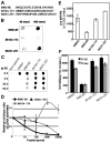
Figure 4. Augmentation of reverse transcription by HIV-1 IN and SIP1 in vitro.
(A, B) The in vitro RT assay was carried out with 35 fmoles of HIV-1 RT (GST-RTp66) in the absence or presence of different amounts of full-length rIN (WT-IN). The truncated form of IN (IN1–70) that lack SIP1 binding domains was also tested in apparel as a control. The amount of HIV-1 cDNA was measured by real-time PCR using the HIV-1 R/U5 (A) or R/gag (B) primer pair . Significant augmentation (p<0.05) by WT-IN in the levels of HIV-1 cDNA compared with the level without IN (0 fmole) was indicated by asterisks. (C) The in vitro RT assay was carried out as described in (A) in either the absence or presence of 3.5 moles of WT-IN, together with different amounts of rSIP1. (D) The in vitro RT assay was carried out with 3.5 fmoles of WT-IN (black bar) or IN1–70 (slash bar), in either the absence (−) or presence (+) of 50 fmoles of rSIP1. (E) The oligo(dT) cellulose resin was incubated with in vitro transcribed HIV-1 RNA for 1 h. After incubation, 10 µg of GST-RT in either the absence or presence of His-IN or rSIP1 was added and incubated for 2 h. After four washes with the IN storage buffer, precipitates were analyzed by immunoblot analysis using an anti-GST antibody.
Figure 5
Figure 5. IN-derived peptide inhibits reverse transcription in vitro and in vivo.
(A) Amino acid sequences of IN-derived peptides are indicated. (B) 40 or 400 nmol of each peptide bound to nitrocellulose filter was incubated with rSIP1 (5 µg). The rSIP1 bound to the peptide was detected using an anti-SIP1 antibody. (C) His-IN (10 µg) was bound to nitrocellulose and reacted with 5 µg of rSIP1 in the presence of each peptide at the indicated concentration. The rSIP1 bound to the peptide was detected as described in (B). (D) The in vitro reverse transcription assay was performed with 35 fmol of RT, 3.5 fmol of His-IN, and 50 fmol of rSIP1 in either the absence (control) or presence of IN-derived peptide at the indicated concentration. The values (mean±SE) plotted are the levels of HIV-1 cDNA relative to that the control, taken as 1.0. (E, F) PMA-stimulated THP-1 cells were treated with 100 µM of IN-derived peptide for 16 h. Cells were infected with HIVNL43-luc/VSV-G pseudotypes in the presence of 100 µM of IN-derived peptide for 6 h. At 24 h post-infection, levels of viral gene expression in cells were determined by measuring the luciferase activity (E) and the levels of viral cDNA synthesis for early (R/U5, black bar) and late (R/gag, slash bar) products (F) were determined as described in Figure 4.

References

    1. Cullen BR. Journey to the center of the cell. Cell. 2001;105:697–700. - PubMed
    1. Goff SP. Intracellular trafficking of retroviral genomes during the early phase of infection: viral exploitation of cellular pathways. J Gene Med. 2001;3:517–528. - PubMed
    1. Katz RA, Skalka AM. The retroviral enzymes. Annu Rev Biochem. 1994;63:133–173. - PubMed
    1. Kalpana GV, Marmon S, Wang W, Crabtree GR, Goff SP. Binding and stimulation of HIV-1 integrase by a human homolog of yeast transcription factor SNF5. Science. 1994;266:2002–2006. - PubMed
    1. Cherepanov P, Maertens G, Proost P, Devreese B, Van Beeumen J, et al. HIV-1 integrase forms stable tetramers and associates with LEDGF/p75 protein in human cells. J Biol Chem. 2003;278:372–381. - PubMed

Publication types