. 2009 Nov 13;4(11):e7809.
doi: 10.1371/journal.pone.0007809.

Epigenetic signatures associated with different levels of differentiation potential in human stem cells

Affiliations

Epigenetic signatures associated with different levels of differentiation potential in human stem cells

Pablo Aranda et al. PLoS One. .

Abstract

Background: The therapeutic use of multipotent stem cells depends on their differentiation potential, which has been shown to be variable for different populations. These differences are likely to be the result of key changes in their epigenetic profiles.

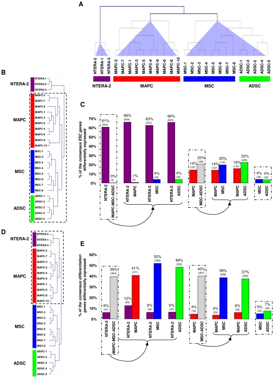
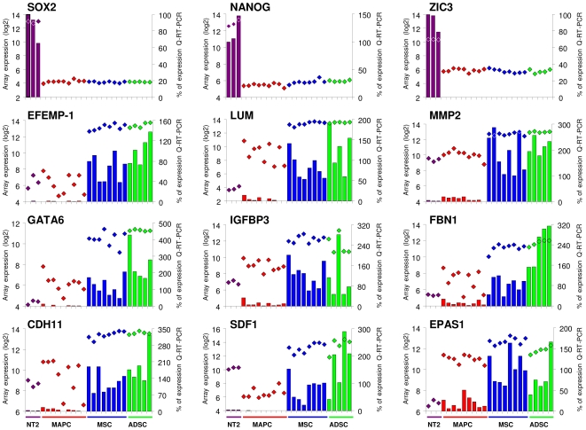
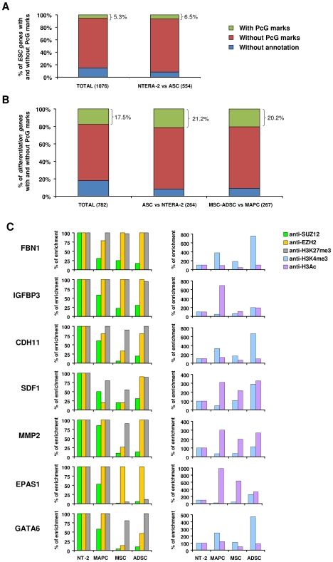
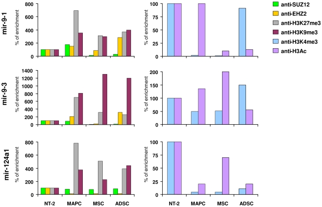
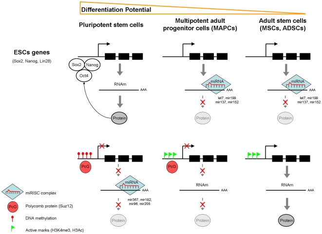
Methodology/principal findings: to address this issue, we have investigated the levels of epigenetic regulation in well characterized populations of pluripotent embryonic stem cells (ESC) and multipotent adult stem cells (ASC) at the trancriptome, methylome, histone modification and microRNA levels. Differences in gene expression profiles allowed classification of stem cells into three separate populations including ESC, multipotent adult progenitor cells (MAPC) and mesenchymal stromal cells (MSC). The analysis of the PcG repressive marks, histone modifications and gene promoter methylation of differentiation and pluripotency genes demonstrated that stem cell populations with a wider differentiation potential (ESC and MAPC) showed stronger representation of epigenetic repressive marks in differentiation genes and that this epigenetic signature was progressively lost with restriction of stem cell potential. Our analysis of microRNA established specific microRNA signatures suggesting specific microRNAs involved in regulation of pluripotent and differentiation genes.

Conclusions/significance: Our study leads us to propose a model where the level of epigenetic regulation, as a combination of DNA methylation and histone modification marks, at differentiation genes defines degrees of differentiation potential from progenitor and multipotent stem cells to pluripotent stem cells.


Conflict of interest statement

Competing Interests: The authors have declared that no competing interests exist.

Figures

Figure 1
Figure 1. Hierarchical clustering and supervised analysis of consensus ESC genes and differentiation genes.
Dendrogram of hierarchical cluster analysis based on the expression of all genes (A), of the consensus ESC genes (B) and consensus differentiation genes (D) included in the HG-U133 Plus 2.0 chip. Hierarchical clustering (Euclidian distance) was performed with the TIGR MeV v. 2.2 program. Analysis of the differential expression of consensus ESC genes (C) and consensus differentiation genes (E) between the populations of stem cells using the significant analysis of microarrays (SAM) algorithm. Values are shown as the percentage of genes that belong to the consensus ESC or consensus differentiation gene lists that are differentially expressed in each group after comparison with SAM. The number of deregulated genes is indicated in parentheses. Only deregulated genes with FC>2 were considered.
Figure 2
Figure 2. Expression of genes from the consensus ESC and differentiation genes lists in NTERA-2, MAPC, MSC and ADSC cells by Q-RT-PCR.
Gene expression was measured using the relative standard curve method. GAPDH was used as a housekeeping control. Expression values of each sample from Affymetrix arrays (logarithmic scale: rhombuses) and from Q-RT-PCR analysis (percentages: bars) are shown for each gene. Q-RT-PCR results are expressed on the secondary vertical axis as a % of expression relative to NTERA-2 for consensus ESC genes, and to MSC for consensus differentiation genes. Each bar represents the value of a different cell line.
Figure 3
Figure 3. Consensus ESC and differentiation genes with predicted Polycomb group marks, and regulation of differentiation genes by PcG proteins in stem cells.
A, Bar graph displaying the percentage of genes targeted by PcG marks among the consensus ESC gene list and among the ESC genes upregulated in NTERA-2 cells relative to ASC. B, Bar graph displaying the percentage of genes targeted by PcG marks among the consensus differentiation gene list, the differentiation genes upregulated in ASC relative to NTERA-2 cells, and differentiation genes upregulated in MSC and ADSC versus MAPC. The numbers in brackets represent the total number of genes. C, Occupancy of EZH2, SUZ12, H3K27m3, H3K4me3 and H3 acetylated, assessed by Q-ChIP-PCR, in the promoters of differentiation genes. Enrichment is presented as a percentage relative to NTERA-2 (100%).
Figure 4
Figure 4. Methylation and expression of genes hypomethylated in ASC in comparison with NTERA-2 cells.
A, Differences of expression between ASC and NTERA-2 (blue bars) and promoter methylation in ASC (black diamonds) and NTERA-2 (red squares) are presented for each gene. The differences of expression, according to Affymetrix data, are shown as fold change (FC) in log2. The methylation data are average BeadArray values. B, Bisulfite sequencing of promoter regions of COL1A2, HOXA9 and SERPINE1 in NTERA-2 and ASC cells. Promoter schematic description (black) and the location of the CpGs (blue) examined are presented for each gene. The CpG dinucleotides (clear box: unmethylated, filled box: methylated) of five clones and the BeadArray methylation values±standard deviation are shown for each sample. C, Expression of COL1A2, HOXA9 and SERPINE1 from Affymetrix arrays (colored rhombuses) and quantitative Q-RT-PCR (colored bars).
Figure 5
Figure 5. Expression of miRNAs and their putative ESC and differentiation target genes.
Expression of ESC genes -NANOG, LIN28 and ZIC3- and differentiation genes -DCN and COL1A2- (dashed lines, log2, left vertical axis) and expression of miRNAs (colored bars, log10, right vertical axis) predicted to regulate expression of these genes were analyzed by Q-RT-PCR in each cell line.
Figure 6
Figure 6. Quantitative-ChIP assay performed on miR-9-1, miR9-3 and -miR124a1.
Antibodies specific to SUZ12, EZH2, H3K27me3, H3K9me3, H3K4me3 and H3 acetylated were used for ChIP, and then the miR-9-1, miR-9-3 and miR-124a1 levels were determined using Q-RT-PCR. Enrichment is measured as percentage relative to NTERA-2 cells (100%).
Figure 7
Figure 7. Model of epigenetic regulation of ESC and differentiation genes in different populations of human stem cells.
Genes implicated in pluripotency (Oct4, Sox2, Nanog) are transcriptionally active in ESC due to the lack of repressive marks as well as by their own potential to transactivate their own transcription favoring the greater differentiation potential of ESC. Differentiation genes, on the contrary are silenced on ESC due to several epigenetic mechanisms that include expression of miRNA, DNA promoter methylation and repressive histone marks that all together cooperate to induce down-regulation of differentiation genes. A decrease in the differentiation potential of ASC (MAPC, MSC and ADSC) is mediated by the silencing of pluripotency genes downregulated through the expression of certain miRNA. The presence of PcG proteins (particularly SUZ12) on the promoters of differentiation genes but the lack of methylation on MAPC could explain their reduced expression. Finally, the lack of PcG marks along with the increase in open chromatin marks in the promoters of differentiation genes would explain the greater expression of these genes as well as the more restricted differentiation potential of ASC (MSC and ADSC).

Similar articles

Cited by

References

    1. Yu J, Thomson JA. Pluripotent stem cell lines. Genes Dev. 2008;22:1987–1997. - PMC - PubMed
    1. Hoffman LM, Carpenter MK. Characterization and culture of human embryonic stem cells. Nat Biotechnol. 2005;23:699–708. - PubMed
    1. Bibikova M, Laurent LC, Ren B, Loring JF, Fan JB. Unraveling epigenetic regulation in embryonic stem cells. Cell Stem Cell. 2008;2:123–134. - PubMed
    1. Bernstein BE, Mikkelsen TS, Xie X, Kamal M, Huebert DJ, et al. A bivalent chromatin structure marks key developmental genes in embryonic stem cells. Cell. 2006;125:315–326. - PubMed
    1. Pan G, Tian S, Nie J, Yang C, Ruotti V, et al. Whole-genome analysis of histone H3 lysine 4 and lysine 27 methylation in human embryonic stem cells. Cell Stem Cell. 2007;1:299–312. - PubMed

Publication types