. 2009 Nov 13;4(11):e7810.
doi: 10.1371/journal.pone.0007810.

Detection of B. anthracis spores and vegetative cells with the same monoclonal antibodies

Affiliations

Detection of B. anthracis spores and vegetative cells with the same monoclonal antibodies

Dian-Bing Wang et al. PLoS One. .

Abstract

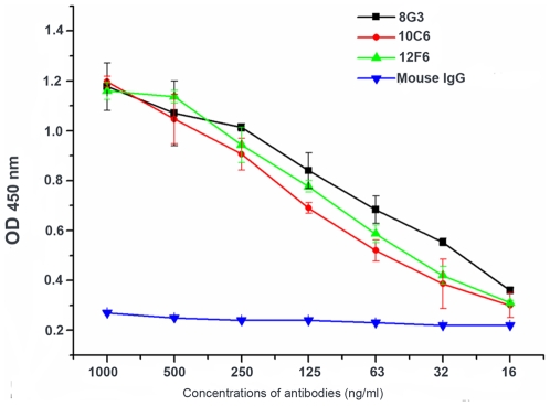
Bacillus anthracis, the causative agent of anthrax disease, could be used as a biothreat reagent. It is vital to develop a rapid, convenient method to detect B. anthracis. In the current study, three high affinity and specificity monoclonal antibodies (mAbs, designated 8G3, 10C6 and 12F6) have been obtained using fully washed B. anthracis spores as an immunogen. These mAbs, confirmed to direct against EA1 protein, can recognize the surface of B. anthracis spores and intact vegetative cells with high affinity and species-specificity. EA1 has been well known as a major S-layer component of B. anthracis vegetative cells, and it also persistently exists in the spore preparations and bind tightly to the spore surfaces even after rigorous washing. Therefore, these mAbs can be used to build a new and rapid immunoassay for detection of both life forms of B. anthracis, either vegetative cells or spores.


Conflict of interest statement

Competing Interests: The authors have declared that no competing interests exist.

Figures

Figure 1
Figure 1. Images captured by AFM analysis of B. anthracis spores.
Topography images (left) and amplitude images (right) of the spores were collected in tapping mode. A: Unwashed spores. B: Extensively washed spores.
Figure 2
Figure 2. The specificity and reactivity of mAbs with B. anthracis spores.
A: Reaction of the mAbs with a series of Bacillus spores, as detected by indirect ELISA. B: Reactions of different concentrations of mAbs with B. anthracis spores by indirect ELISA. The experiment was repeated three times.
Figure 3
Figure 3. Transmission electron micrographs.
A: B. anthracis exosporium fragments and the B. anthracis spores after sonication. B: Scale bars: 500 nm.
Figure 4
Figure 4. Identification of the mAbs' target proteins.
A: The protein profiles of the fully washed B. anthracis exosporium following SDS-PAGE (Lane 1) and immunoblotting with mAb 8G3 (Lane 2), 10C6 (Lane 3), 12F6 (Lane 4) and mouse IgG (Lane 5). B: Mass spectrometric analysis of the target protein, which was determined to be EA1.
Figure 5
Figure 5. The reactivity of the mAbs with purified EA1 protein.
Reactions of a range of concentrations of the mAbs with the 4 µg/ml EA1 protein using indirect ELISA. The experiment was repeated three times.
Figure 6
Figure 6. The specificity and reactivity of the mAbs with B. anthracis vegetative cells.
Reactions of the mAbs with a series of vegetative cells, as detected by sandwich ELISA.
Figure 7
Figure 7. Representative confocal microscopy images of 8G3 binding to fully washed B. anthracis spores (top) and vegetative cells (bottom).
The secondary antibodies were R-phycoerythrin-conjugated goat anti-mouse IgG. (red) or FITC-conjugated goat anti-mouse IgG (green). The corresponding phase contrast images (A and D) and fluorescence images (B and E) were merged to show overlap (C and F). Scale bars: spores, 5 µm; vegetative cells, 10 µm.
Figure 8
Figure 8. Comparison of the protein profiles of spores and debris.
Proteins were separated on a 10% Tricine gel and visualized with silver staining. Lane 1, unwashed spores (Preparation 1); Lane 2, spores washed three times with ultrapure water (Preparation 2); Lane 3, spores prepared by Renografin purification (Preparation 3); Lane 4, fully washed spores (Preparation 4); Lane 5, debris in the supernatant of centrifuged spores after Renografin purification. (The staining time of Preparation 3, 4 and 5 was longer than that of Preparation 1 and 2, in order that more bands of each sample could be visualized clearly).

References

    1. Green BD, Battisti L, Koehler TM, Thorne CB, Ivins BE. Demonstration of a capsule plasmid in Bacillus anthracis. Infect Immun. 1985;49:291–297. - PMC - PubMed
    1. Mikesell P, Ivins BE, Ristroph JD, Dreier TM. Evidence for plasmid-mediated toxin production in Bacillus anthracis. Infect Immun. 1983;39:371–376. - PMC - PubMed
    1. Nicholson WL, Munakata N, Horneck G, Melosh HJ, Setlow P. Resistance of Bacillus endospores to extreme terrestrial and extraterrestrial environments. Microbiol Mol Biol Rev. 2000;64:548–572. - PMC - PubMed
    1. Ash C, Collins MD. Comparative analysis of 23S ribosomal RNA gene sequences of Bacillus anthracis and emetic Bacillus cereus determined by PCR-direct sequencing. FEMS Microbiol Lett. 1992;73:75–80. - PubMed
    1. Daffonchio D, Cherif A, Borin S. Homoduplex and heteroduplex polymorphisms of the amplified ribosomal 16S–23S internal transcribed spacers describe genetic relationships in the “Bacillus cereus group”. Appl Environ Microbiol. 2000;66:5460–5468. - PMC - PubMed

Publication types