. 2009 Nov 4:2:25.
doi: 10.3389/neuro.02.025.2009. eCollection 2009.

Localization of glycine receptors in the human forebrain, brainstem, and cervical spinal cord: an immunohistochemical review

Affiliations

Localization of glycine receptors in the human forebrain, brainstem, and cervical spinal cord: an immunohistochemical review

Kristin Baer et al. Front Mol Neurosci. .

Abstract

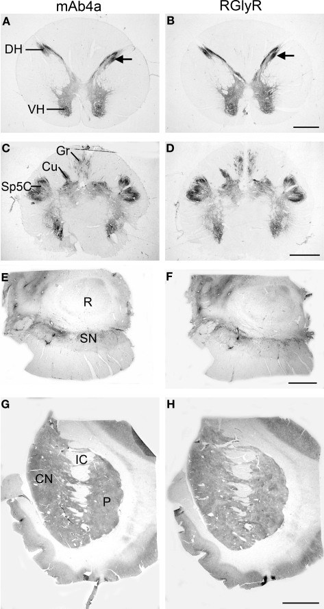
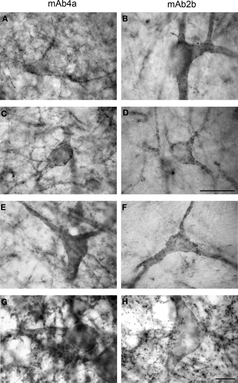
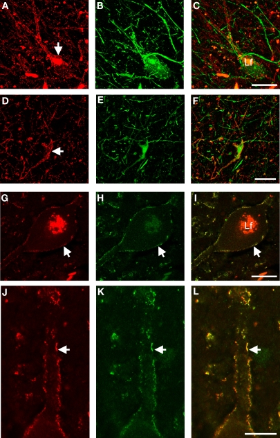
Inhibitory neurotransmitter receptors for glycine (GlyR) are heteropentameric chloride ion channels that are comprised of four functional subunits, alpha1-3 and beta and that facilitate fast-response, inhibitory neurotransmission in the mammalian brain and spinal cord. We have investigated the distribution of GlyRs in the human forebrain, brainstem, and cervical spinal cord using immunohistochemistry at light and confocal laser scanning microscopy levels. This review will summarize the present knowledge on the GlyR distribution in the human brain using our established immunohistochemical techniques. The results of our immunohistochemical labeling studies demonstrated GlyR immunoreactivity (IR) throughout the human basal ganglia, substantia nigra, various pontine regions, rostral medulla oblongata and the cervical spinal cord present an intense and abundant punctate IR along the membranes of the neuronal soma and dendrites. This work is part of a systematic study of inhibitory neurotransmitter receptor distribution in the human CNS, and provides a basis for additional detailed physiological and pharmacological studies on the inter-relationship of GlyR, GABA(A)R and gephyrin in the human brain. This basic mapping exercise, we believe, will provide important baselines for the testing of future pharmacotherapies and drug regimes that modulate neuroinhibitory systems. These findings provide new information for understanding the complexity of glycinergic functions in the human brain, which will translate into the contribution of inhibitory mechanisms in paroxysmal disorders and neurodegenerative diseases such as Epilepsy, Huntington's and Parkinson's Disease and Motor Neuron Disease.

Keywords: glycine receptor; human brain; immunohistochemistry.


Figures

Figure 1
Figure 1
Regional localization of glycine receptors at the cervical level of the human spinal cord (A,B), pyramidal decussation in the lower medulla oblongata (C,D), substantia nigra (E,F) and basal ganglia at the level of the caudate-putamen (G,H) visualized by DAB staining of adjacent sections using monoclonal antibody mAb4a (A,C,E,G) and rabbit polyclonal antibody AB5052 (B,D,F,H). (A,B) Light macroscopic images of sections of the first cervical segment of the spinal cord stained for GlyR with mAb4a (A) and AB5052 (B) antibody showing especially intense labeling in lamina II of the dorsal horn (DH) and moderate to high levels of GlyR-IR in the gray matter of the ventral horn (VH) and dorsal horn. (C,D) Macroscopic images of sections of the pyramidal decussation stained for GlyR with mAb4a (C) and AB5052 (D) antibody revealing patchy GlyR-IR in the gracile nucleus (Gr) and the cuneate nucleus (Cu) and intense GlyR-IR in the spinal trigeminal nucleus (Sp5C). (E,F) Macroscopic images of sections of the substantia nigra (SN) stained for GlyR with mAb4a (G) and AB5052 (H) antibody showing high levels of GlyR-IR in the regions of the substantia nigra pars compacta and the substantia nigra pars reticulata. (G,H) Macroscopic images of sections of the basal ganglia at the level of the caudate-putamen stained for GlyR with mAb4a (E) and AB5052 (F) antibody suggesting a heterogeneous distribution of GlyR in the caudate nucleus (CN) and putamen (P) separated by the internal capsule (IC). Scale bar = 2 mm in (A,B); 2.5 mm in (C,D); 0.5 cm in (E–H).
Figure 2
Figure 2
Glycine receptor labeling on neurons in the human midbrain and medulla oblongata visualized by DAB staining of adjacent sections using monoclonal antibody mAb4a (A,C,E,G) and monoclonal antibody mAb2b (B,D,F,H). (A,B) Neurons in the periaqueductal grey area of the midbrain showing high levels of punctate GlyR-IR along their soma and processes, revealing similar IR patterns using mAb4a (A) or mAb2b (B). (C,D) Neurons in the superior colliculus of the midbrain immunoreactive for mAb4a (C) and mAb2b (D) demonstrating GlyR-IR outlining the membranes of soma and processes. (E,F) Neurons in the magnocellular region of the reticular formation in the medulla oblongata immunoreactive for mAb4a (E) and mAb2b (F) revealing GlyR-IR along the neuronal soma and processes. (G,H) High levels of immunoreactive GlyR are present on neurons in the hypoglossal nucleus of the upper medulla stained for mAb4a (G) and mAb2b (H). Scale bar = 50 μm in (D) applies to (C–F); 10 μm in H [applies to (A,B,G,H)].
Figure 3
Figure 3
High-resolution confocal laser scanning microscopy images of neurons in the substantia nigra pars reticulata (A–C), putamen (D–F), and first cervical segment of the spinal cord (G–L) of the human CNS double-labeled for GlyR (left column) and other cell markers (middle column) with combined images shown in the right column. (A–C) Neuron in the substantia nigra pars reticulata is labeled red for GlyR [mAb4a in (A), arrow] and immunopositive for rabbit anti-parvalbumin [green in (B)], green unlabeled parvalbumin processes in the picture resemble axon bundles, combined images in (C). (D–F) Neuron in the putamen that is outlined with GlyR-IR [mAb4a, red in (D), arrow] is labeled with rabbit anti-calretinin immunoreactivity [green in (E)], combined images in (F). (G–I) Large neuron in the spinal cord that is outlined with rabbit anti-GlyR immunoreactivity [red in (G)] is labeled with mouse anti-gephyrin immunoreactivity [green in (H)], combined images in (I). Note the high degree of overlap of both markers, exemplified by the arrow highlighting the colocalized IR patterns. (J–L) High magnification of a process of a large motoneuron in the cervical spinal cord that shows punctate GlyR-IR [red in (J)] and punctate gephyrin-IR [green in (K)] demonstrating the high degree of colocalization of the GlyR and gephyrin IR patterns [yellow in (L)]. Lf in (C) and (I) indicates lipofuchsin autofluorescence in the cell body. Scale bar = 25 μm in (C) [applies to (A–C)], (F) [applies to (D–F)]; (G) [applies to (G–I)], and (J) [applies to (J–L)].

References

    1. Adams J. C. (1981). Heavy metal intensification of DAB-based HRP reaction product. J. Histochem. Cytochem. 29, 775. - PubMed
    1. Altschuler R. A., Betz H., Parakkal M. H., Reeks K. A., Wenthold R. J. (1986). Identification of glycinergic synapses in the cochlear nucleus through immunocytochemical localization of the postsynaptic receptor. Brain Res. 369, 316–320 10.1016/0006-8993(86)90542-1 - DOI - PubMed
    1. Alvarez F. J., Dewey D. E., Harrington D. A., Fyffe R. E. (1997). Cell-type specific organization of glycine receptor clusters in the mammalian spinal cord. J. Comp. Neurol. 379, 150–170 10.1002/(SICI)1096-9861(19970303)379:1<150::AID-CNE10>3.0.CO;2-T - DOI - PubMed
    1. Andrew M., Owen M. J. (1997). Hyperekplexia: abnormal startle response due to glycine receptor mutations. Br. J. Psychiatry 170, 106–108 10.1192/bjp.170.2.106 - DOI - PubMed
    1. Baer K., Waldvogel H. J., During M. J., Snell R. G., Faull R. L. M., Rees M. I. (2003). Association of gephyrin and glycine receptors in the human brainstem and spinal cord: an immunohistochemical analysis. Neuroscience 122, 773–784 10.1016/S0306-4522(03)00543-8 - DOI - PubMed

LinkOut - more resources