. 2009 Nov 13;4(11):e7814.
doi: 10.1371/journal.pone.0007814.

Dermatitis and aging-related barrier dysfunction in transgenic mice overexpressing an epidermal-targeted claudin 6 tail deletion mutant

Affiliations

Dermatitis and aging-related barrier dysfunction in transgenic mice overexpressing an epidermal-targeted claudin 6 tail deletion mutant

Tammy-Claire Troy et al. PLoS One. .

Abstract

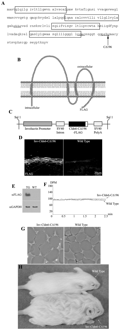
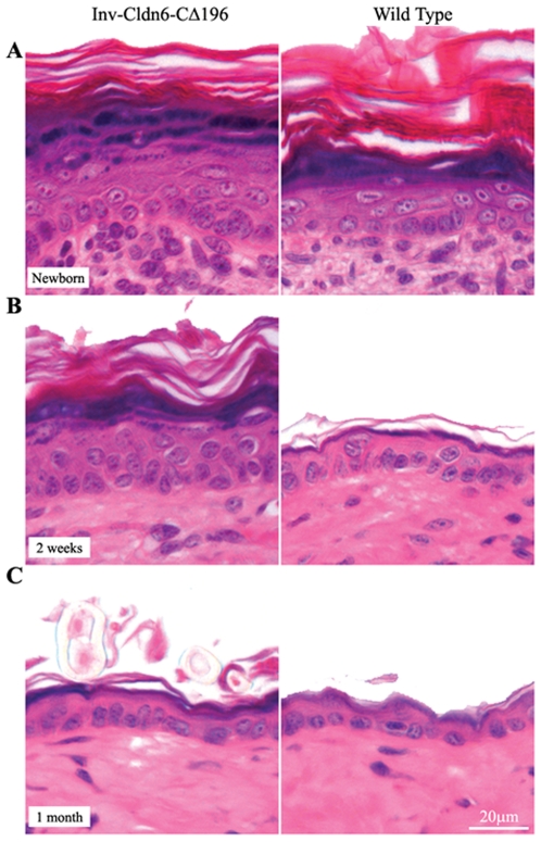
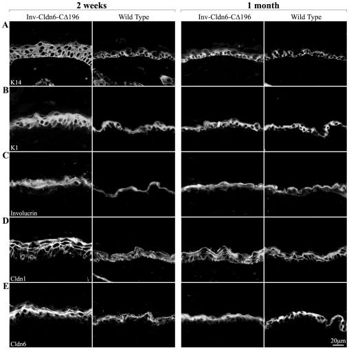
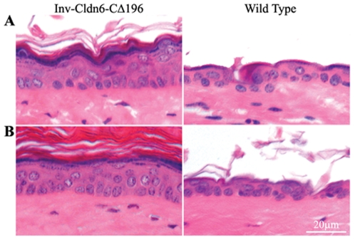
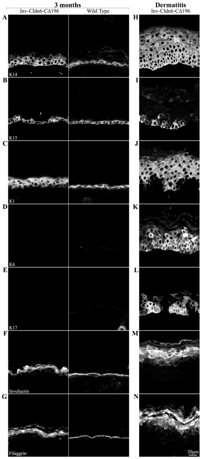
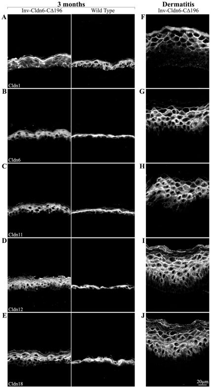
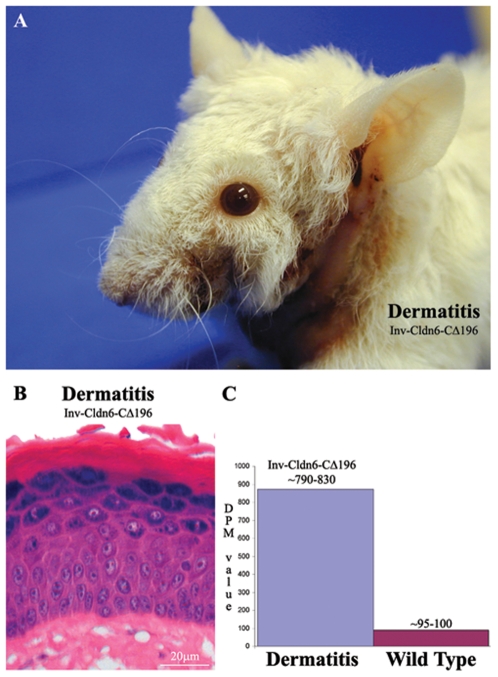
The barrier function of the skin protects the mammalian body against infection, dehydration, UV irradiation and temperature fluctuation. Barrier function is reduced with the skin's intrinsic aging process, however the molecular mechanisms involved are unknown. We previously demonstrated that Claudin (Cldn)-containing tight junctions (TJs) are essential in the development of the epidermis and that transgenic mice overexpressing Cldn6 in the suprabasal layers of the epidermis undergo a perturbed terminal differentiation program characterized in part by reduced barrier function. To dissect further the mechanisms by which Cldn6 acts during epithelial differentiation, we overexpressed a Cldn6 cytoplasmic tail deletion mutant in the suprabasal compartment of the transgenic mouse epidermis. Although there were no gross phenotypic abnormalities at birth, subtle epidermal anomalies were present that disappeared by one month of age, indicative of a robust injury response. However, with aging, epidermal changes with eventual chronic dermatitis appeared with a concomitant barrier dysfunction manifested in increased trans-epidermal water loss. Immunohistochemical analysis revealed aberrant suprabasal Cldn localization with marked down-regulation of Cldn1. Both the proliferative and terminal differentiation compartments were perturbed as evidenced by mislocalization of multiple epidermal markers. These results suggest that the normally robust injury response mechanism of the epidermis is lost in the aging Involucrin-Cldn6-CDelta196 transgenic epidermis, and provide a model for evaluation of aging-related skin changes.


Conflict of interest statement

Competing Interests: The authors have declared that no competing interests exist.

Figures

Figure 1
Figure 1. Inv-Cldn6-CΔ196 transgenic mice.
The Cldn6 protein sequence is shown; transmembrane-spanning regions are encased within boxes, the CXXC motifs are underlined, and the truncation site is indicated with an arrow (A). The Inv-Cldn6-CΔ196 mutant was created by deleting the C-terminal half of the cytoplasmic tail domain of Cldn6 after amino acid 196 (B). The Inv promoter was use to drive transgene expression to the suprabasal compartment of the transgenic mouse epidermis, where TJs are localized (C). Transgene expression was confirmed to be restricted to the upper spinous and granular layers of the epidermis as visualized by immunohistochemistry using anti-FLAG antibodies (D) and immunoblotting confirmed a ∼19.5kDa band corresponding to the transgene product in skin samples from transgenic (TG) but not wild type (WT) skin samples using anti-GAPDH as a loading control (E). Trans-epidermal water loss measurements confirmed no skin barrier dysfunction in the Inv-Cldn6-CΔ196 transgenic mice at birth (F). This was further supported by evaluation of cornified envelopes; which were characterized by a rigid shape and uniform size comparable to that of the wild type (G). Inv-Cldn6-CΔ196 mice were easily identifiable by their frizzed and lackluster coat appearance as compared to the wild type, a phenotype that persisted throughout life (H).
Figure 2
Figure 2. Histological abnormalities in the Inv-Cldn6-CΔ196 transgenic epidermis.
Histological analysis of newborn (A) Inv-Cldn6-CΔ196 transgenic mice as compared to the wild type revealed a moderately thicker epidermis in transgenic samples, with a thicker stratum corneum, a less-compacted granular layer and an overall expanded suprabasal compartment. At 2-weeks of age (B), the Inv-Cldn6-CΔ196 epidermis remained somewhat less mature and thicker than that of the wild type; however by 1-month of age (C) the transgenic and wild type samples were histologically indistinguishable.
Figure 3
Figure 3. Perturbation of markers of epidermal differentiation.
Immunofluorescence was used to evaluate various markers of epidermal differentiation including K14 (A), K1 (B), involucrin (C), Cldn1 (D) and Cldn6 (E) in Inv-Cldn6-CΔ196 mice as compared to their age-matched wild type counterparts. In each case abnormalities were present in samples from 2-week old (left panel) transgenic mice that were normalized by 1-month of age (right panel). For example, the K14 expression zone was extended into the suprabasal compartment the Inv-Cldn6-CΔ196 epidermis until epidermal thinning was achieved. In addition, broadened K1 and involucrin expression compartments was seen early but normalized by 1-month of age. Sporadic Cldn1-positive basal cells were evident in the 2-week old transgenic epidermis; by 1-month of age Cldn1-positive cells occupied both the basal and suprabasal compartments reminiscent of the wild type. Cldn6, a suprabasal-specific Cldn, was observed in an expanded zone early on, and was normalized by 1-month of age.
Figure 4
Figure 4. Histological evidence of epidermal abnormalities in the aging Inv-Cldn6-CΔ196 transgenic epidermis.
Histological evidence of epidermal abnormalities emerged in aging Inv-Cldn6-CΔ196 transgenic mice after 3 (A) and 6 (B) months as compared to the wild type. These included an expanded stratum corneum, as well as an expanded suprabasal zone and a less compacted granular layer suggesting an intrinsic propensity for injury and inefficient repair that increases with aging in the Inv-Cldn6-CΔ196 epidermis.
Figure 5
Figure 5. Aberrance in markers of epidermal differentiation during aging.
Changes in the epidermal differentiation program of the aging Inv-Cldn6-CΔ196 transgenic epidermis were evaluated by immunofluorescence and compared to their age-matched wild type counterparts (samples from 3-month-old mice are shown – left panel); their expression/localization in dermatitis-affected areas is also indicated (right panel). K14 was expressed throughout the basal and suprabasal compartments in the aging Inv-Cldn6-CΔ196 epidermis (A), while K15 remained restricted to basal cells (B). The zone of K1 expression was expanded in the 3-month-old transgenic epidermis (C); however, neither K6- (D) nor K17-positive (E) cells were observed. An expanded zone of expression, and improper packing, of involucrin (F) and filaggrin (G) suggested perturbations in terminal differentiation program of the aged Inv-Cldn6-CΔ196 epidermis. In the dermatitis-affected areas, K14 (H) expression was expanded far into the suprabasal compartment, while K15 was only sporadically observed (I). A punctate K1 localization was evident throughout the thickened suprabasal zone of the dermatitis-affected Inv-Cldn6-CΔ196 epidermis (J), and K6- (K) and K17-positive (L) basal and suprabasal cells were observed. In addition, involucrin (M) and filaggrin (N) expression compartments were also expanded, with obvious packing defects evident.
Figure 6
Figure 6. Evaluation of Cldns in the aging Inv-Cldn6-CΔ196 transgenic epidermis.
The expression/localization of Cldns was also perturbed in the aging Inv-Cldn6-CΔ196 transgenic epidermis (samples are from 3-month-old mice – left panel); the dermatitis-affected zones are also shown (right panel). Cldn1-positive basal and suprabasal cells were reduced in the aging Inv-Cldn6-CΔ196 epidermis (A). In addition, a shift away from a strictly membrane localization and an expanded zone of suprabasal Cldns was evident - Cldn6 (B), Cldn11 (C), Cldn12 (D) and Cldn18 (E). These abnormalities in were strikingly exacerbated in the dermatitis-affected epidermis. For instance, the basal and lower suprabasal layers were void of Cldn1 localization (F). In addition, Cldn6 (G), Cldn11 (H), Cldn12 (I) and Cldn18 (J) localization was observed throughout the expanded suprabasal zone with membranous localization less evident in the lower suprabasal layers.
Figure 7
Figure 7. Aging-associated barrier dysfunction.
Upon gross observation, obvious signs of dermatitis were evident in aging Inv-Cldn6-CΔ196 transgenic mice, especially in areas subjected to repetitive mechanical stress such as the neck and behind the ears (A). Histological evaluation revealed a significantly thickened epidermis with an increased number of spinous/suprabasal cell layers and cellular disorganization; the upper differentiation layers were also abnormal, with parakeratosis, the prevalent appearance of nuclei, an improperly packed granular layer and a thicker stratum corneum (B). TEWL measurements across the dermatitis-affected transgenic skin confirmed an approximately 8-fold increase in water loss and a barrier deficient phenotype (C).

Similar articles

Cited by

References

    1. Fuchs E, Raghavan S. Getting under the skin of epidermal morphogenesis. Nat Rev Genet. 2002;3:199–209. - PubMed
    1. Fuchs E. Scratching the surface of skin development. Nature. 2007;445:834–842. - PMC - PubMed
    1. Turksen K, Troy TC. Epidermal cell lineage. Biochem Cell Biol. 1998;76:889–898. - PubMed
    1. Pouillot A, Dayan N, Polla AS, Polla LL, Polla BS. The stratum corneum: a double paradox. J Cosmet Dermatol. 2008;7:143–148. - PubMed
    1. Elias PM, Williams ML, Holleran WM, Jiang YJ, Schmuth M. Pathogenesis of permeability barrier abnormalities in the ichthyoses: inherited disorders of lipid metabolism. J Lipid Res. 2008;49:697–714. - PMC - PubMed

Publication types

MeSH terms