. 2010 Feb;231(3):507-21.
doi: 10.1007/s00425-009-1049-5. Epub 2009 Nov 14.

Two novel disaccharides, rutinose and methylrutinose, are involved in carbon metabolism in Datisca glomerata

Affiliations

Two novel disaccharides, rutinose and methylrutinose, are involved in carbon metabolism in Datisca glomerata

Maria Schubert et al. Planta. 2010 Feb.

Erratum in

  • Planta. 2010 Feb;231(3):523

Abstract

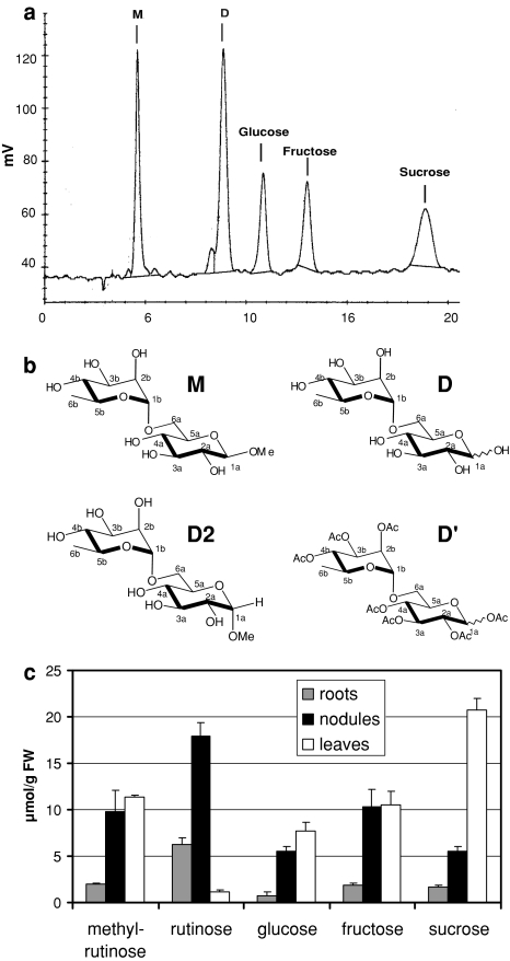
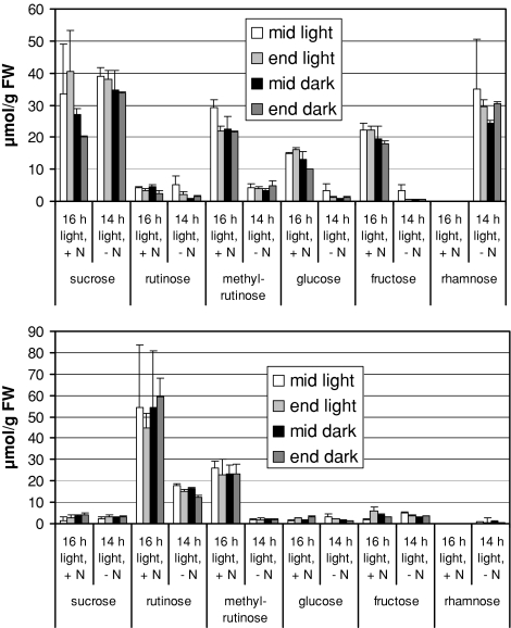
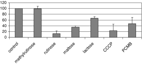
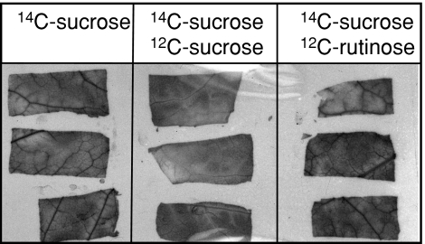
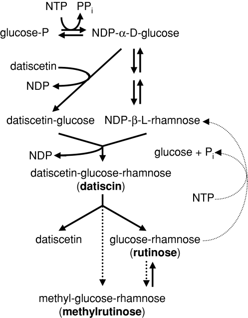
Datisca glomerata forms nitrogen-fixing root nodules in symbiosis with soil actinomycetes from the genus Frankia. Analysis of sugars in roots, nodules and leaves of D. glomerata revealed the presence of two novel compounds that were identified as alpha-L-rhamnopyranoside-(1 --> 6)-D-glucose (rutinose) and alpha-L-rhamnopyranoside-(1 --> 6)-1-O-beta-D-methylglucose (methylrutinose). Rutinose has been found previously as a/the glycoside part of several flavonoid glycosides, e.g. rutin, also of datiscin, the main flavonoid of Datisca cannabina, but had not been reported as free sugar. Time course analyses suggest that both rutinose and methylrutinose might play a role in transient carbon storage in sink organs and, to a lesser extent, in source leaves. Their concentrations show that they can accumulate in the vacuole. Rutinose, but not methylrutinose, was accepted as a substrate by the tonoplast disaccharide transporter SUT4 from Arabidopsis. In vivo (14)C-labeling and the study of uptake of exogenous sucrose and rutinose from the leaf apoplast showed that neither rutinose nor methylrutinose appreciably participate in phloem translocation of carbon from source to sink organs, despite rutinose being found in the apoplast at significant levels. A model for sugar metabolism in D. glomerata is presented.


Figures

Fig. 1
Fig. 1
Identification and distribution of rutinose and methylrutinose. a A sugar HPLC chromatogram of a nodule extract showing two peaks (labeled with “D” and “M”, respectively) that could not be identified via comparison with standards. b Chemical structure of rutinose (D) and methylrutinose (M). The product of the chemical acetylation of D (D′) showed that the connection between the monosaccharides was 1b (rhamnose) to 6a (glucose). Chemical methylation of rutinose led to a α-glycosidic bond in the 1a position (D2), in contrast to the β-glycosidic bond in the natural substance (M). c Sugar contents of roots (gray bars), nodules (black bars) and leaves (white bars) of greenhouse-grown D. glomerata (150 μmol photons m−2 s−1). Average values from three parallel greenhouse-grown series (150 μmol photons m−2 s−1) are shown
Fig. 2
Fig. 2
Time course of sugar contents (μmol/g fresh weight) in roots and leaves of D. glomerata. Plants were grown in aerated hydroponic culture at 16 h light, 8 h dark or 14 h light, 10 h dark, respectively, and samples (in duplicate) were taken for determination of sugar content in the middle of the light phase, 30 min before the end of the light phase, in the middle of the dark phase and 30 min before the end of the dark phase. The plants grown at 16 h light received 5 mM KNO3 each week. The KNO3 in the growth medium of the plants grown at 14 h light had not been replenished since 3 weeks at the harvesting date
Fig. 3
Fig. 3
Electron micrographs of D. glomerata minor veins. CC companion cell, SE sieve element, PP phloem parenchyma, X xylem element, XP xylem parenchyma. a Overview of a minor vein. b Upper sieve element-companion cell complex on an adjacent section. c Detail of the left companion cell and adjacent mesophyll cell shows two plasmodesmata (arrows). The size bars denote 5 μm (a), 2.5 μm (b) and 1 μm (c), respectively
Fig. 4
Fig. 4
Substrate specificity of AtSUT4 in yeast cells. Uptake of 3 mM 14C-sucrose by strain SEY2102 (Emr et al. 1983) containing AtSUT4 in pDR196 (Weise et al. 2000) was analyzed in the presence of inhibitors and of competing sugars. The sulfhydryl group inhibitor PCMBS and the uncoupler CCCP were used as inhibitors and added to a final concentration of 50 μM. Uptake of 3 mM 14C-sucrose was also analyzed in the presence of competing non-labeled sugars added at a threefold excess (final concentrations of 9 mM). Each bar represents results from at least three independent experiments. Standard deviations are given
Fig. 5
Fig. 5
Uptake of 14C-sucrose by D. glomerata source leaves. D. glomerata leaves were fed buffered 14C-sucrose solution either without additives, or supplied with 20 mM 12C-sucrose or 20 mM 12C-rutinose, respectively. After washing, parts of the laminae containing minor veins were excised, dried and autoradiographed
Fig. 6
Fig. 6
Hypothetical model for (methyl-) rutinose metabolism in D. glomerata. Solid arrows denote reactions shown in D. glomerata or in other plants, dashed arrows show hypothetical reactions. NDP-β-l-rhamnose is synthesized via NDP-α- d-glucose. Sequential glycosylation of the flavonol datiscetin with nucleotide sugars leads first to datiscetin-glucose (datiscanin; Zapesochnaya et al. 1982b) and then to datiscetin-rutinoside (datiscin). A flavonol 3-O-β-heterodisaccharidase cleaves datiscin into datiscetin and rutinose or catalyzes a transglycosylation yielding datiscetin and methylrutinose; this reaction can take place in extracts from roots, nodules and leaves. In all those organs, methylrutinose can be demethylated by a β-glucosidase of unknown subcellular localization, yielding rutinose. It is not clear whether rutinose can be methylated to methylrutinose, or whether methylrutinose can only be formed from datiscin by transglycosylation. Since no α-rhamnosidase activity was detected and since rhamnose never was found to accumulate in sink organs of D. glomerata to any significant extent, in contrast with other sugars, rutinose cleavage is postulated to yield NDP-β-l-rhamnose

Similar articles

Cited by

References

    1. Altschul SF, Gish W, Miller W, Myers EW, Lipman DJ. Basic local alignment search tool. J Mol Biol. 1990;215:304–310. - PubMed
    1. Aoki N, Hirose T, Scofield GN, Whitfeld PR, Furbank RT. The sucrose transporter gene family in rice. Plant Cell Physiol. 2003;44:223–232. doi: 10.1093/pcp/pcg030. - DOI - PubMed
    1. Avigad G. Sucrose and other disaccharides. In: Loewus FA, Tanner W, editors. Plant carbohydrates I: intracellular carbohydrates. Encyclopedia of plant physiology, volume 13A. Berlin: Springer; 1982. pp. 217–347.
    1. Bar-Peled M, Lewinsohn E, Fluhr R, Gressel J. UDP-rhamnose:flavanone-7-O-glucoside-2′α″-O-rhamnosyltransferase. Purification and characterization of an enzyme catalyzing the production of bitter compounds in Citrus. J Biol Chem. 1991;226:20953–20959. - PubMed
    1. Baumgertel A, Grimm R, Eisenbeiß W, Kreis W. Purification and characterization of a flavonol 3-O-β-heterodisaccharidase from the dried herb of Fagopyrum esculentum Moench. Phytochemistry. 2003;64:411–418. doi: 10.1016/S0031-9422(03)00418-7. - DOI - PubMed

Publication types

LinkOut - more resources