Establishment and characterization of an osteopontin-null cutaneous squamous cell carcinoma cell line
- PMID: 19915934
- PMCID: PMC3664068
- DOI: 10.1007/s11626-009-9248-8
Establishment and characterization of an osteopontin-null cutaneous squamous cell carcinoma cell line
Abstract
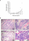
Osteopontin (OPN) is a secreted glycoprotein implicated to function in cancer development and metastasis. Although elevated expression of OPN are observed in cancer cells of various types, in some cases, only the cells in the stromal region surrounding the tumor express OPN, suggesting distinct functional roles for this protein derived from host cells and from cancer cells. To provide a model for addressing the functions and mechanisms of host-derived OPN in cancer progression and metastasis, a cutaneous squamous cell carcinoma cell line (ONSC) that lacks the OPN gene, Spp1, was established. This line of cells was derived from a squamous cell carcinoma that developed in a female, OPN-null mouse subjected to two-stage skin carcinogenesis. Morphologically, ONSC cells resemble epithelial cells, and they express the epithelial markers, K1, K14, and p63, as confirmed by immunohistochemical analyses. Genomic analyses indicate the presence of mutated H-Ras and p53 genes. ONSC cells form colonies in soft agar and, subcutaneously injected into athymic nude mice, develop into squamous cell carcinomas that metastasize to the lungs. Lacking OPN expression, these squamous cell carcinoma cells provide a model to address the function of host OPN in the context of cancer progression and metastasis.
Figures


Similar articles
-
Osteopontin facilitates ultraviolet B-induced squamous cell carcinoma development.J Dermatol Sci. 2014 Aug;75(2):121-32. doi: 10.1016/j.jdermsci.2014.05.002. Epub 2014 May 21. J Dermatol Sci. 2014. PMID: 24888687 Free PMC article.
-
Host-derived osteopontin maintains an acute inflammatory response to suppress early progression of extrinsic cancer cells.Int J Cancer. 2012 Jul 15;131(2):322-33. doi: 10.1002/ijc.26359. Epub 2011 Oct 20. Int J Cancer. 2012. PMID: 21826648 Free PMC article.
-
Distinct roles of osteopontin in host defense activity and tumor survival during squamous cell carcinoma progression in vivo.Cancer Res. 1998 Nov 15;58(22):5206-15. Cancer Res. 1998. PMID: 9823334
-
Role of osteopontin in cellular signaling and metastatic phenotype.Front Biosci. 2008 May 1;13:4276-84. doi: 10.2741/3004. Front Biosci. 2008. PMID: 18508510 Review.
-
Transcriptional regulation of osteopontin and the metastatic phenotype: evidence for a Ras-activated enhancer in the human OPN promoter.Clin Exp Metastasis. 2003;20(1):77-84. doi: 10.1023/a:1022550721404. Clin Exp Metastasis. 2003. PMID: 12650610 Review.
Cited by
-
Osteopontin facilitates ultraviolet B-induced squamous cell carcinoma development.J Dermatol Sci. 2014 Aug;75(2):121-32. doi: 10.1016/j.jdermsci.2014.05.002. Epub 2014 May 21. J Dermatol Sci. 2014. PMID: 24888687 Free PMC article.
-
Host-derived osteopontin maintains an acute inflammatory response to suppress early progression of extrinsic cancer cells.Int J Cancer. 2012 Jul 15;131(2):322-33. doi: 10.1002/ijc.26359. Epub 2011 Oct 20. Int J Cancer. 2012. PMID: 21826648 Free PMC article.
-
Repeated exposure of the oral mucosa over 12 months with cold plasma is not carcinogenic in mice.Sci Rep. 2021 Oct 19;11(1):20672. doi: 10.1038/s41598-021-99924-3. Sci Rep. 2021. PMID: 34667240 Free PMC article.
References
-
- Chang PL, Prince CW. 1alpha,25-dihydroxyvitamin D3 stimulates synthesis and secretion of nonphosphorylated osteopontin (secreted phosphoprotein 1) in mouse JB6 epidermal cells. Cancer Res. 1991;51:2144–2150. - PubMed
-
- Chang PL, Tucker MA, Hicks PH, Prince CW. Novel protein kinase C isoforms and mitogen-activated kinase kinase mediate phorbol ester-induced osteopontin expression. Intl. J. Biochem. Cell Biol. 2002;34:1142–1151. - PubMed
-
- Colburn NH. Tumor promoter produces anchorage independence in mouse epidermal cells by an induction mechanism. Carcinogenesis. 1980;1:951–954. - PubMed
-
- Crawford HC, Matrisian LM, Liaw L. Distinct roles of osteopontin in host defense activity and tumor survival during squamous cell carcinoma progression in vivo. Cancer Res. 1998;58:5206–5215. - PubMed
Publication types
MeSH terms
Substances
Grants and funding
LinkOut - more resources
Full Text Sources
Medical
Research Materials
Miscellaneous

