

Structural and functional characterization of the integral membrane protein VDAC-1 in lipid bilayer nanodiscs
- PMID: 19916553
- PMCID: PMC2793270
- DOI: 10.1021/ja907918r
Structural and functional characterization of the integral membrane protein VDAC-1 in lipid bilayer nanodiscs
Abstract
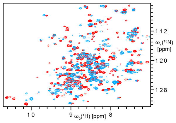
Biophysical studies of membrane proteins are often impeded by the requirement for a membrane mimicking environment. Detergent micelles are the most common choice, but the denaturing properties make them unsatisfactory for studies of many membrane proteins and their interactions. In the present work, we explore phospholipid bilayer nanodiscs as membrane mimics and employ electron microscopy and solution NMR spectroscopy to characterize the structure and function of the human voltage dependent anion channel (VDAC-1) as an example of a polytopic integral membrane protein. Electron microscopy reveals the formation of VDAC-1 multimers, an observation that is consistent with results obtained in native mitochondrial outer membranes. High-resolution NMR spectroscopy demonstrates a well folded VDAC-1 protein and native NADH binding functionality. The observed chemical shift changes upon addition of the native ligand NADH to nanodisc-embedded VDAC-1 resemble those of micelle-embedded VDAC-1, indicating a similar structure and function in the two membrane-mimicking environments. Overall, the ability to study integral membrane proteins at atomic resolution with solution NMR in phospholipid bilayers, rather than in detergent micelles, offers exciting novel possibilities to approach the biophysical properties of membrane proteins under nondenaturing conditions, which makes this technology particular suitable for protein-protein interactions and other functional studies.
Figures


References
-
- Seddon AM, Curnow P, Booth PJ. Biochim Biophys Acta. 2004;1666:105–17. - PubMed
-
- Prosser RS, Evanics F, Kitevski JL, Al-Abdul-Wahid MS. Biochemistry. 2006;45:8453–65. - PubMed
-
- Caffrey M. J Struct Biol. 2003;142:108–32. - PubMed
-
- Marcotte I, Auger M. Concepts Magn Reson Part A. 2005;24A:17–37.
-
- Jonas A. Methods Enzymol. 1986;128:553–82. - PubMed
Publication types
MeSH terms
Substances
Grants and funding
LinkOut - more resources
Full Text Sources
Other Literature Sources
