. 2009 Dec;158(8):1904-10.
doi: 10.1111/j.1476-5381.2009.00482.x.

Potentiation by WIN 55,212-2 of GABA-activated currents in rat trigeminal ganglion neurones

Affiliations

Potentiation by WIN 55,212-2 of GABA-activated currents in rat trigeminal ganglion neurones

Zhi-Wang Li et al. Br J Pharmacol. 2009 Dec.

Abstract

Background and purpose: Although both natural and synthetic cannabinoid compounds have been shown to exert an antinociceptive effect on acute and persistent pain, the anatomical locus of the target of cannabinoid-induced analgesia has not been fully elucidated. Here, we investigated the effects of the cannabinoid agonist WIN 55,212-2 on GABA-activated currents (I(GABA)) in rat primary sensory neurones.

Experimental approach: In the present study, experiments were performed on neurones freshly isolated from rat trigeminal ganglion (TG) by using whole-cell patch clamp and repatch techniques.

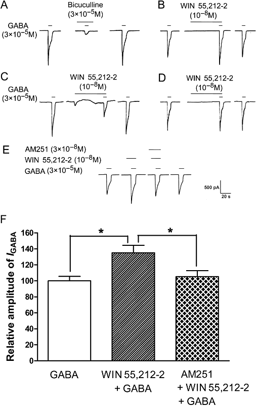
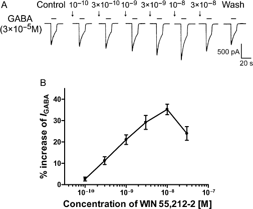
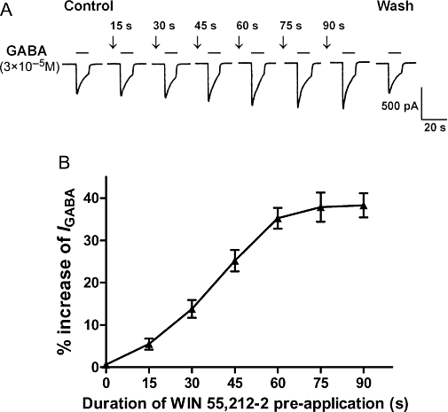
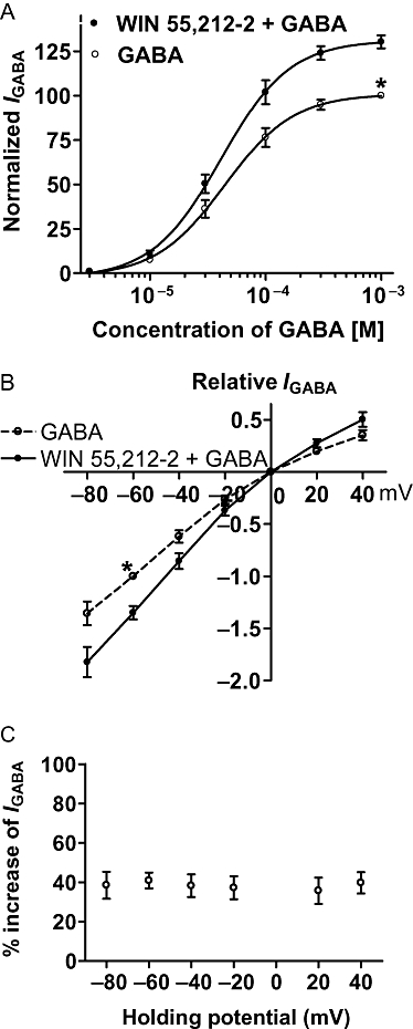
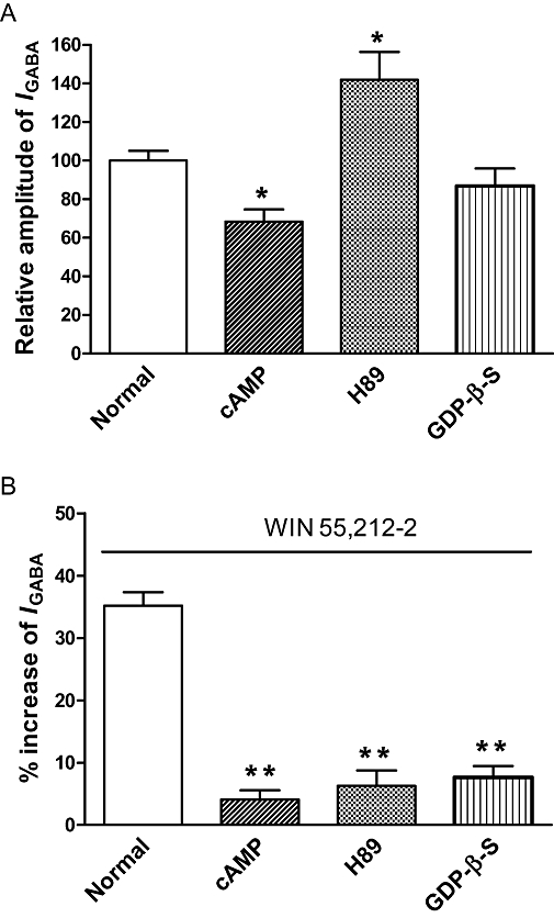
Key results: GABA-evoked inward currents were potentiated by pretreatment with WIN 55,212-2 in a concentration-dependent manner (10(-10)-10(-8) M). WIN 55,212-2 shifted the GABA concentration-response curve upwards, with an increase of 30.3 +/- 3.7% in the maximal current response but with no significant change in the EC(50) (agonist concentration producing a half-maximal response) value. WIN 55,212-2 potentiated the responses to GABA in a manner independent of holding potential and in the absence of any change in the reversal potential of the current. This potentiation of I(GABA) induced by WIN 55,212-2 was almost completely blocked by AM 251 (3 x 10(-8) M), a CB(1) receptor antagonist, and, using the repatch technique, was found to be abolished after intracellular dialysis with the protein kinase A (PKA) activator cAMP or the PKA inhibitor H89.

Conclusions and implications: The potentiation by WIN 55,212-2 of I(GABA) in primary sensory neurones may help to elucidate the mechanism underlying the modulation of analgesia by cannabinoids in the spinal dorsal horn.


Figures

Figure 1
Figure 1
Modulatory effects of WIN 55,212-2 on GABA-activated currents in rat trigeminal ganglion (TG) neurones. (A) The inward current evoked by 3 × 10⁻⁵ M GABA could be blocked by the GABAA receptor antagonist bicuculline. Pre-application of WIN 55,212-2 (10−8 M) exerts an enhancing (B), inhibitory (C) and no effect (D) on GABA-activated currents in rat TG neurones. (E) Current traces demonstrating that the CB1 receptor antagonist AM 251 blocked the potentiation by WIN 55,212-2 of GABA-activated currents. (F) Shows the inhibitory effect of AM 251 (3 × 10⁻⁸ M) on WIN 55,212-2 (10−8 M)-induced potentiation of GABA (3 × 10⁻⁵ M)-activated currents. *P < 0.05, one way analysis of variance followed by Bonferroni's post hoc test, n= 7.
Figure 2
Figure 2
Concentration-dependent potentiation of GABA-activated currents by WIN 55,212-2. (A) Sequential current-traces illustrating the potentiation of IGABA induced by different concentrations of WIN 55,212-2(10−10–3 × 10⁻⁸ M) on a single TG neurone. (B) Shows that IGABA increased step by step on increasing the concentration of WIN 55,212-2 from 10−10 to 10−8 M; at the higher concentration, the increase of IGABA reached its peak value; beyond that, IGABA gradually decreased. The pretreatment time for WIN 55,212-2 was 60 s. Each point represents the mean ± SEM of 7–9 neurones.
Figure 3
Figure 3
Effect of the duration of the pre-application of WIN 55,212-2 on its potentiation of GABA-activated currents. (A) Current traces demonstrating that the increase of IGABA induced by WIN 55,212-2 (10−8 M) is related to the duration of its pre-application from 15 to 90 s. (B) Shows that the potentiation of IGABA (3 × 10⁻⁵ M) by WIN 55,212-2 (10−8 M) was enhanced when the duration of the pre-application was increased from 15 to 90 s. Note that, at a pre-application duration of between 75 and 90 s, the potentiation of the GABA-activated current reached its maximum value. Each point represents the mean ± SEM of 6–9 neurones.
Figure 4
Figure 4
Concentration–response and current–voltage (I-V) relationships for GABA with or without the pre-application of WIN 55,212-2. (A) The concentration–response curves for GABA with or without WIN 55,212-2 (10−8 M) pre-application. Each point represents the mean ± SEM of 8–10 neurones. All GABA-induced currents were normalized to the response induced by 10−3 M GABA applied alone (marked with asterisk). (B) The I-V curves for GABA (3 × 10⁻⁵ M)-activated current with and without WIN 55,212-2 (10−8 M) pre-appplication. All current values from the same cell were normalized to the current response induced by 10−3 M GABA applied alone (marked with asterisk). Each point represents the mean ± SEM of 6–8 neurones. The experiment was carried out by using recording pipettes filled with CsCl containing internal solution. (C) Increase of IGABA (%) evoked by WIN 55,212-2 (10−8 M) at different holding potentials. Each point represents the mean ± SEM of 6–8 neurones.
Figure 5
Figure 5
G-protein coupling and intracellular phosphorylation are shown to be involved in the potentiation of GABA-activated currents by WIN 55,212-2. (A) Intracellular dialysis of cAMP, an activator of PKA, significantly decreased IGABA. H89, a PKA inhibitor, significantly increased IGABA, while dialysis of guanosine 5=-O-(2-thiodiphosphate)(GDP)-β-S, a non-hydrolysable GDP analogue, had no effect on IGABA. *P < 0.05, post hoc Bonferroni's test, compared with normal internal solution, n= 7/each column. (B) Intracellular dialysis of cAMP, H89 and GDP-β-S abolished the enhancing effect of WIN 55,212-2 (10−8 M) on IGABA. The duration of the pre-application of WIN 55,512-2 was 30 s. **P < 0.01, post hoc Bonferroni's test, compared with normal internal solution, n= 7/each column.

Similar articles

Cited by

References

    1. Agarwal N, Pacher P, Tegeder I, Amaya F, Constantin CE, Brenner GJ, et al. Cannabinoids mediate analgesia largely via peripheral type 1 cannabinoid receptors in nociceptors. Nat Neurosci. 2007;10:870–879. - PMC - PubMed
    1. Ahluwalia J, Urban L, Capogna M, Bevan S, Nagy I. Cannabinoid 1 receptors are expressed in nociceptive primary sensory neurons. Neuroscience. 2000;100:685–688. - PubMed
    1. Bridges D, Rice AS, Egertová M, Elphick MR, Winter J, Michael GJ. Localisation of cannabinoid receptor 1 in rat dorsal root ganglion using in situ hybridisation and immunohistochemistry. Neuroscience. 2003;119:803–812. - PubMed
    1. Calignano A, La Rana G, Giuffrida A, Piomelli D. Control of pain initiation by endogenous cannabinoids. Nature. 1998;394:277–281. - PubMed
    1. Chen QX, Stelzer A, Kay AR, Wong RK. GABAA receptor function is regulated by phosphorylation in acutely dissociated guinea-pig hippocampal neurons. J Physiol. 1990;420:207–221. - PMC - PubMed

MeSH terms