

Analysis of expressed sequence tags and identification of genes encoding cell-wall-degrading enzymes from the fungivorous nematode Aphelenchus avenae
- PMID: 19917084
- PMCID: PMC2784482
- DOI: 10.1186/1471-2164-10-525
Analysis of expressed sequence tags and identification of genes encoding cell-wall-degrading enzymes from the fungivorous nematode Aphelenchus avenae
Abstract
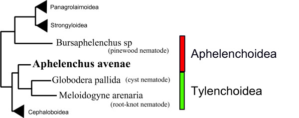
Background: The fungivorus nematode, Aphelenchus avenae is widespread in soil and is found in association with decaying plant material. This nematode is also found in association with plants but its ability to cause plant disease remains largely undetermined. The taxonomic position and intermediate lifestyle of A. avenae make it an important model for studying the evolution of plant parasitism within the Nematoda. In addition, the exceptional capacity of this nematode to survive desiccation makes it an important system for study of anhydrobiosis. Expressed sequence tag (EST) analysis may therefore be useful in providing an initial insight into the poorly understood genetic background of A. avenae.
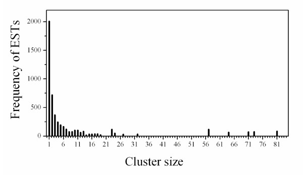
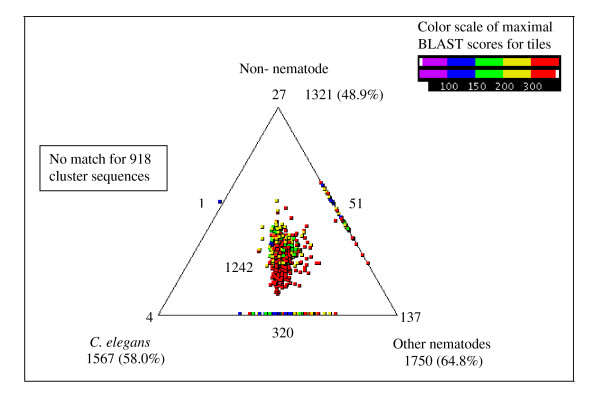
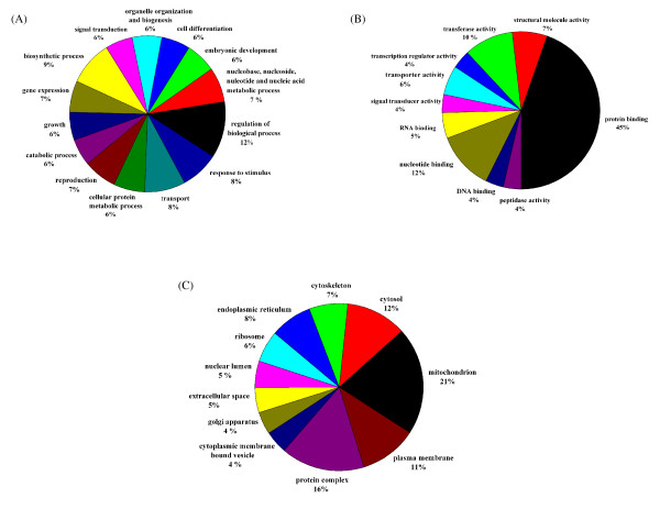
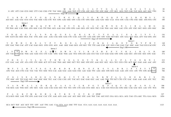
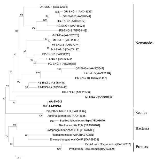
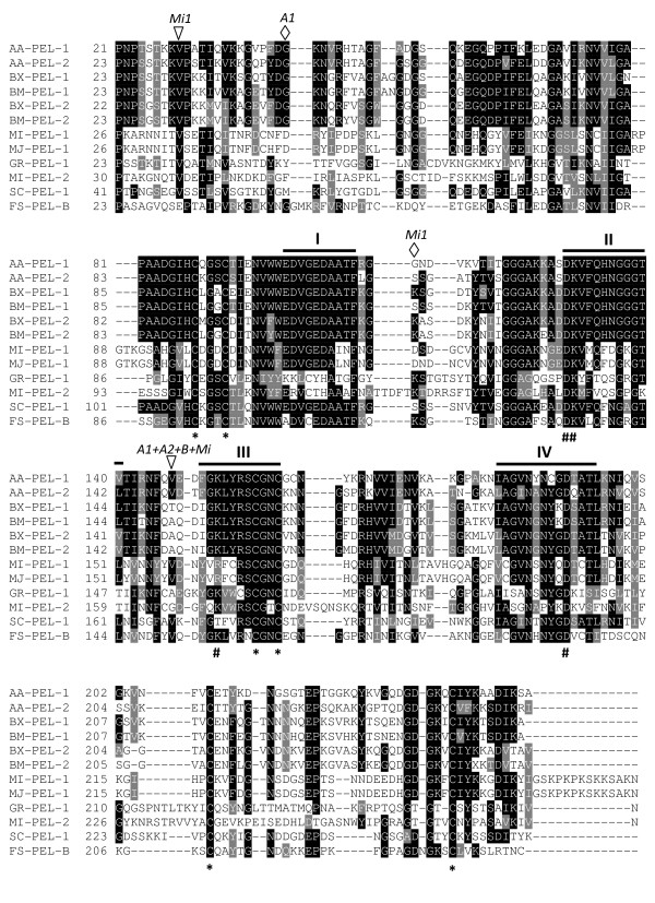
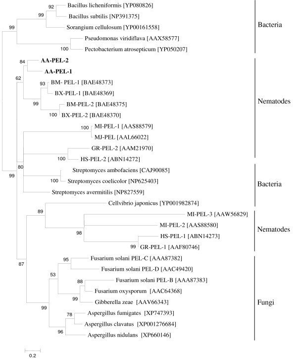
Results: We present the generation, analysis and annotation of over 5,000 ESTs from a mixed-stage A. avenae cDNA library. Clustering of 5,076 high-quality ESTs resulted in a set of 2,700 non-redundant sequences comprising 695 contigs and 2,005 singletons. Comparative analyses indicated that 1,567 (58.0%) of the cluster sequences had homologues in Caenorhabditis elegans, 1,750 (64.8%) in other nematodes, 1,321(48.9%) in organisms other than nematodes, and 862 (31.9%) had no significant match to any sequence in current protein or nucleotide databases. In addition, 1,100 (40.7%) of the sequences were functionally classified using Gene Ontology (GO) hierarchy. Similarity searches of the cluster sequences identified a set of genes with significant homology to genes encoding enzymes that degrade plant or fungal cell walls. The full length sequences of two genes encoding glycosyl hydrolase family 5 (GHF5) cellulases and two pectate lyase genes encoding polysaccharide lyase family 3 (PL3) proteins were identified and characterized.
Conclusion: We have described at least 2,214 putative genes from A. avenae and identified a set of genes encoding a range of cell-wall-degrading enzymes. This EST dataset represents a starting point for studies in a number of different fundamental and applied areas. The presence of genes encoding a battery of cell-wall-degrading enzymes in A. avenae and their similarities with genes from other plant parasitic nematodes suggest that this nematode can act not only as a fungal feeder but also a plant parasite. Further studies on genes encoding cell-wall-degrading enzymes in A. avenae will accelerate our understanding of the complex evolutionary histories of plant parasitism and the use of genes obtained by horizontal gene transfer from prokaryotes.
Figures
Similar articles
-
Expression profiling and cross-species RNA interference (RNAi) of desiccation-induced transcripts in the anhydrobiotic nematode Aphelenchus avenae.BMC Mol Biol. 2010 Jan 19;11:6. doi: 10.1186/1471-2199-11-6. BMC Mol Biol. 2010. PMID: 20085654 Free PMC article.
-
Desiccation survival in an Antarctic nematode: molecular analysis using expressed sequenced tags.BMC Genomics. 2009 Feb 9;10:69. doi: 10.1186/1471-2164-10-69. BMC Genomics. 2009. PMID: 19203352 Free PMC article.
-
Rather than by direct acquisition via lateral gene transfer, GHF5 cellulases were passed on from early Pratylenchidae to root-knot and cyst nematodes.BMC Evol Biol. 2012 Nov 21;12:221. doi: 10.1186/1471-2148-12-221. BMC Evol Biol. 2012. PMID: 23171084 Free PMC article.
-
Role of horizontal gene transfer in the evolution of plant parasitism among nematodes.Methods Mol Biol. 2009;532:517-35. doi: 10.1007/978-1-60327-853-9_30. Methods Mol Biol. 2009. PMID: 19271205 Review.
-
Horizontal gene transfer in nematodes: a catalyst for plant parasitism?Mol Plant Microbe Interact. 2011 Aug;24(8):879-87. doi: 10.1094/MPMI-03-11-0055. Mol Plant Microbe Interact. 2011. PMID: 21539433 Review.
Cited by
-
Assessment of the behaviour and survival of nematodes under low oxygen concentrations.PLoS One. 2018 May 14;13(5):e0197122. doi: 10.1371/journal.pone.0197122. eCollection 2018. PLoS One. 2018. PMID: 29758056 Free PMC article.
-
Insights into metazoan evolution from Alvinella pompejana cDNAs.BMC Genomics. 2010 Nov 16;11:634. doi: 10.1186/1471-2164-11-634. BMC Genomics. 2010. PMID: 21080938 Free PMC article.
-
Horizontal gene transfer of acetyltransferases, invertases and chorismate mutases from different bacteria to diverse recipients.BMC Evol Biol. 2016 Apr 12;16:74. doi: 10.1186/s12862-016-0651-y. BMC Evol Biol. 2016. PMID: 27068610 Free PMC article.
-
Fungi-Nematode Interactions: Diversity, Ecology, and Biocontrol Prospects in Agriculture.J Fungi (Basel). 2020 Oct 4;6(4):206. doi: 10.3390/jof6040206. J Fungi (Basel). 2020. PMID: 33020457 Free PMC article. Review.
-
Detoxification-related gene expression accompanies anhydrobiosis in the foliar nematode (Aphelenchoides fragariae).J Nematol. 2020;52:1-12. doi: 10.21307/jofnem-2020-047. J Nematol. 2020. PMID: 32449331 Free PMC article.
References
-
- Gilleard JS. The use of Caenorhabditis elegans in parasitic nematode research. Parasitology. 2004;128(Suppl 1):S49–S70. - PubMed
-
- De Ley P, Blaxter M. In: Biology of Nematodes. Donald LL, editor. London, UK: Taylor and Francis; 2002. Systematic position and phylogeny; pp. 1–30.
-
- Barker KR. On the disease reduction and reproduction of the nematode Aphelenchus avenae on isolates of Rhizoctonia solani. Plant Dis Reptr. 1964;48:428–432.
-
- Klink JW, Barker KR. Effect of Aphelenchus avenae on the survival and pathogenic activity of root-rotting fungi. Phytopathology. 1968;58:228–232.
Publication types
MeSH terms
Substances
Associated data
- Actions
- Actions
- Actions
- Actions
- Actions
- Actions
- Actions
- Actions
- Actions
- Actions
LinkOut - more resources
Full Text Sources
Research Materials
