. 2009 Nov 18;97(10):2684-92.
doi: 10.1016/j.bpj.2009.09.002.

Anthropomorphizing the mouse cardiac action potential via a novel dynamic clamp method

Affiliations

Anthropomorphizing the mouse cardiac action potential via a novel dynamic clamp method

Rebecca C Ahrens-Nicklas et al. Biophys J. .

Abstract

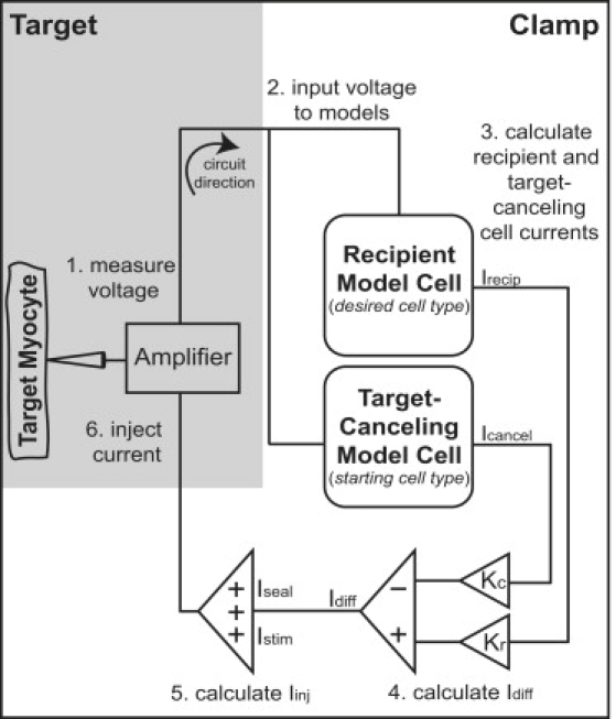
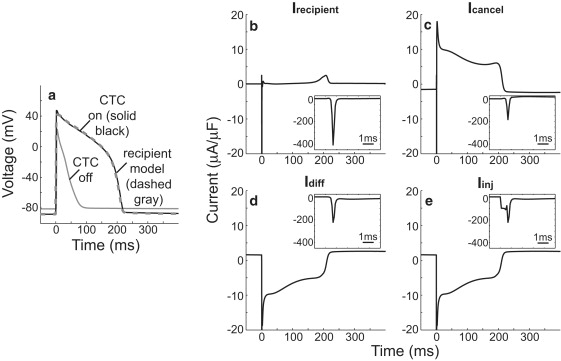
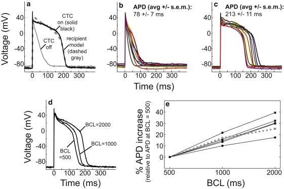
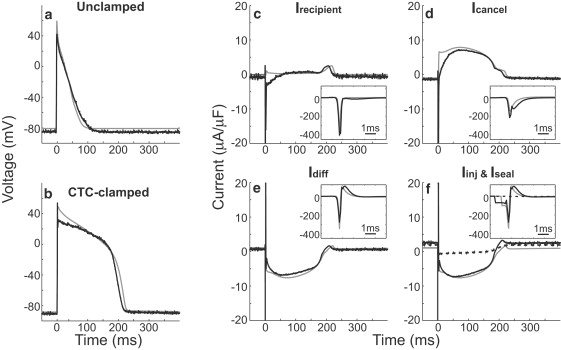
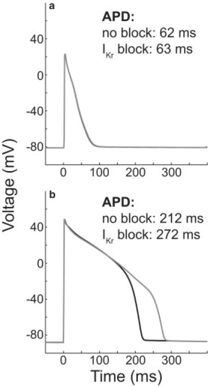
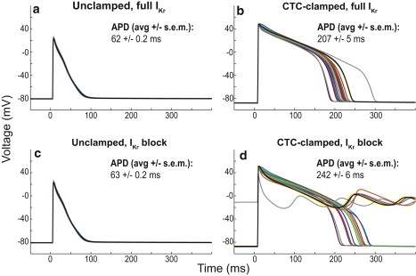
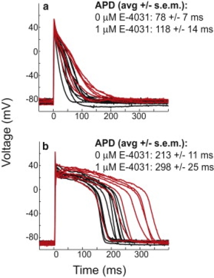
Interspecies differences can limit the translational value of excitable cells isolated from model organisms. It can be difficult to extrapolate from a drug- or mutation-induced phenotype in mice to human pathophysiology because mouse and human cardiac electrodynamics differ greatly. We present a hybrid computational-experimental technique, the cell-type transforming clamp, which is designed to overcome such differences by using a calculated compensatory current to convert the macroscopic electrical behavior of an isolated cell into that of a different cell type. We demonstrate the technique's utility by evaluating drug arrhythmogenicity in murine cardiomyocytes that are transformed to behave like human myocytes. Whereas we use the cell-type transforming clamp in this work to convert between mouse and human electrodynamics, the technique could be adapted to convert between the action potential morphologies of any two cell types of interest.


Figures

Figure 1
Figure 1
Cell-type transforming clamp circuit. An isolated target myocyte is coupled to two computational models (the recipient model cell is the desired cell type, whereas the target-canceling model cell is the same cell type as the target cell). The target cell voltage is measured (step 1) and input to the recipient and target-canceling models (step 2). Each model cell current is calculated and scaled by the ratio of the target cell capacitance to the model cell capacitance (Kc and Kr) (step 3). A difference current, Idiff, is calculated by subtracting the scaled target-canceling model current from the scaled recipient model current (step 4). A stimulus current (Istim) and a current to compensate for leak through the patch-clamp seal (Iseal) are added to the difference current to produce the injected current (Iinj) (step 5), which is injected into the target cell (step 6). The circuit is traversed in real time (at a rate of 10 kHz) to convert the target cell waveform to that of the desired cell type. The experimental (target) portion of the circuit is shaded in gray, whereas the computational (clamp) part of the circuit is white.
Figure 2
Figure 2
CTC can make the waveform of a neonatal mouse model cardiomyocyte humanlike in silico. The CTC was used to clamp an untreated neonatal mouse cardiac myocyte computational model cell (a). The CTC prolonged the short action potential of the unclamped cell (solid gray versus black traces) and induced a phase-3 plateau. The cells were paced at a rate of 1 Hz. The action potential from a statically paced recipient model cell (a, dashed gray) is shown for comparison. The currents responsible for the transition are shown (b–e). Idiff (d) is calculated by subtracting Icancel (c) from Irecipient (b). Iinj results from the addition of a stimulus current to Idiff (e). The insets enlarge the first 6 ms of the action potential to show peak current values.
Figure 3
Figure 3
CTC can anthropomorphize the action potential of real isolated neonatal mouse myocytes in vitro. The CTC was used to clamp an isolated neonatal mouse myocyte (a). The CTC converted the short mouse action potential (gray) into that of the recipient model cell (black). The action potential from a statically paced recipient model cell (a, dashed gray) is shown for comparison. Results from 10 different cells without clamping (b) and during clamping (c) are shown (traces of the same color in b and c correspond to the same cell). Cells were stimulated at 1 Hz. In panels d and e after 20 s with the CTC on in the absence of pacing, cells were stimulated for 20 cycles at 2 Hz, followed by 20 cycles at 1 Hz, and finally 20 cycles at 0.5 Hz. Representative rate-dependent action potential morphologies from one cell are shown (d). Rate-dependent percentage changes in the mean APD for five cells are shown (e). The APD response of the recipient model is shown (e, dotted gray).
Figure 4
Figure 4
In silico and in vitro CTC circuits use similar currents to anthropomorphize a target cell. Traces from in vitro (black) and in silico CTC circuits (gray) are shown. In the in silico circuit, the 80-ms modified Pandit model was used as both the target cell and target-canceling model. Under both unclamped (a) and clamped (b) conditions, the model and real experimental voltages are similar. Irecipient (c), Icancel (d), Idiff (e), and Iinj (f) from both the in silico and in vitro studies are shown. The insets enlarge the first 6 ms of the action potential to show peak current values. The dashed black line of panel f shows the experimental Iseal. Cells were stimulated at 1 Hz. The model cell stimulus was 0.5 ms in duration and 100 μA/μF in magnitude. The experimental cell stimulus was 150% of threshold, 1 ms in duration and 71 μA/μF in magnitude.
Figure 5
Figure 5
CTC reveals the arrhythmogenic potential of IKr-blockade in silico. Model cells in the presence of IKr-blockade (gray traces) demonstrated longer action potentials relative to cells without IKr block (black traces) only during CTC-clamping (b versus a). Cells were stimulated at 1 Hz.
Figure 6
Figure 6
CTC-clamped IKr-blocked myocytes accurately demonstrate drug-induced APD prolongation even in the presence of mismatch between the target and target-canceling model cell in silico. Twenty wild-type and IKr-blocked model variants were created. The original unperturbed Wang and Sobie model was the target-canceling model; therefore, there was mismatch between the target and target-canceling cell in each case. Traces of the same color in panels a–d represent results from the same model variant. In the unclamped cell, there was no major difference between the APDs of blocked and unblocked cells (a versus c). However, in the CTC-clamped models, even in the presence of cell-to-cell variation, the APD-prolonging effects of IKr-block were seen in every model (b versus d). Mismatch between the target and target-canceling cell led to EADs in six out of the 20 models tested. These six traces are examples where model mismatch led to an overestimation of the effects of IKr block. The measured average APD value in the CTC-clamped IKr-blocked cells (242 ms) is artificially short because beats with EADs could not be included in the quantification. Cells were stimulated at 1 Hz for 100 unclamped cycles followed by 20 no-stimulus CTC-clamped cycles and 80 stimulated CTC-clamped cycles. In cells with EADs, full repolarization did not occur before the next stimulus and EADs persisted even after 200 additional CTC-clamped cycles.
Figure 7
Figure 7
CTC reveals the arrhythmogenic potential of the IKr-blocking drug, E-4031. In real isolated cells, IKr-blockade by E-4031 prolonged the unclamped action potential (a, red-treated (n = 7) versus black-untreated (n = 10), two-tailed unpaired t-test, p = 0.015). However, in CTC-clamped real myocytes, E-4031-induced prolongation was increased by 45 ms (b, red-treated versus black-untreated, two-tailed unpaired t-test, p = 0.003). Cells were stimulated at 1 Hz.

References

    1. Salama G., London B. Mouse models of long QT syndrome. J. Physiol. 2007;578:43–53. - PMC - PubMed
    1. Nerbonne J.M. Studying cardiac arrhythmias in the mouse—a reasonable model for probing mechanisms? Trends Cardiovasc. Med. 2004;14:83–93. - PubMed
    1. Doerr T., Denger R., Trautwein W. Calcium currents in single SA nodal cells of the rabbit heart studied with action potential clamp. Pflugers Arch. 1989;413:599–603. - PubMed
    1. Wilders R. Dynamic clamp: a powerful tool in cardiac electrophysiology. J. Physiol. 2006;576:349–359. - PMC - PubMed
    1. Berecki G., Zegers J.G., Verkerk A.O., Bhuiyan Z.A., de Jonge B. HERG channel (dys)function revealed by dynamic action potential clamp technique. Biophys. J. 2005;88:566–578. - PMC - PubMed

Publication types

MeSH terms

LinkOut - more resources