. 2009 Nov 18;97(10):2761-70.
doi: 10.1016/j.bpj.2009.08.054.

Mechanosensitive closed-closed transitions in large membrane proteins: osmoprotection and tension damping

Affiliations

Mechanosensitive closed-closed transitions in large membrane proteins: osmoprotection and tension damping

Pierre-Alexandre Boucher et al. Biophys J. .

Abstract

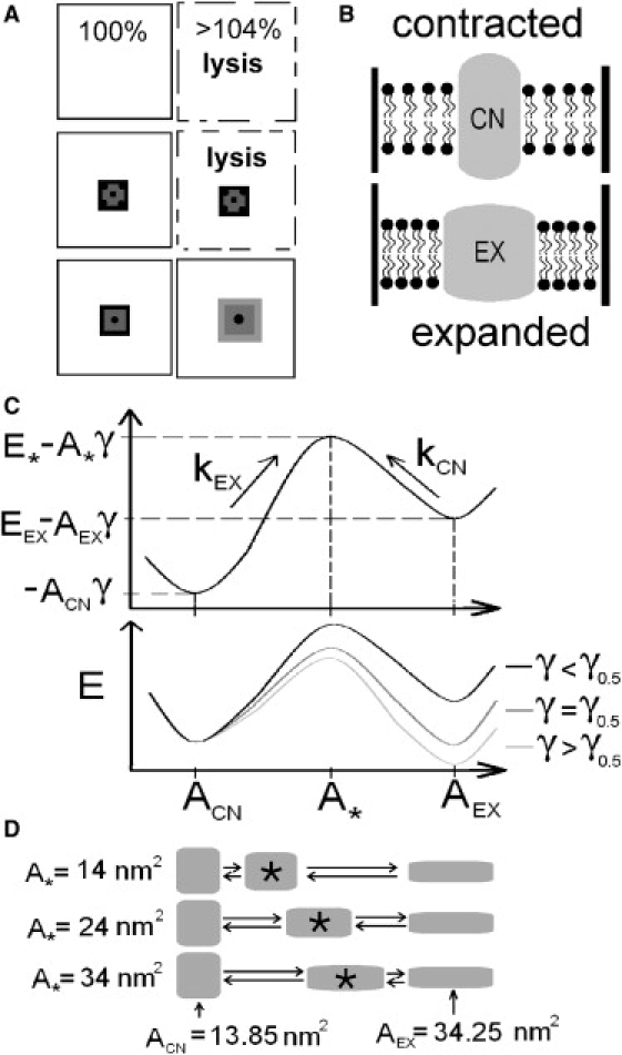
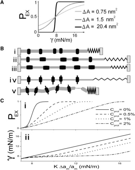
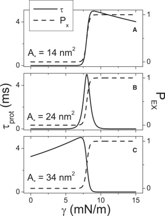
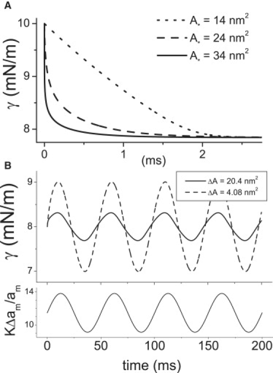
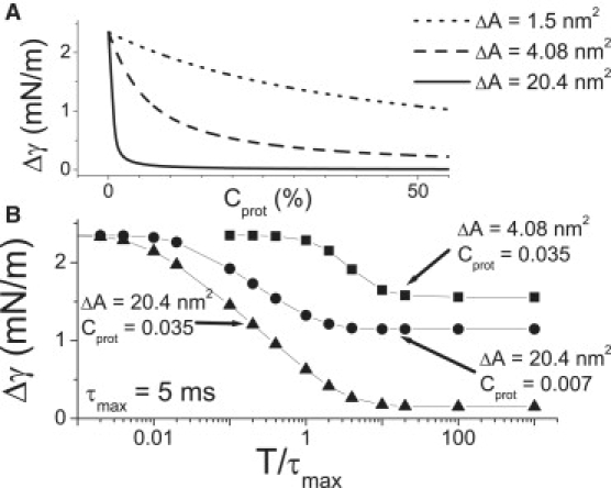
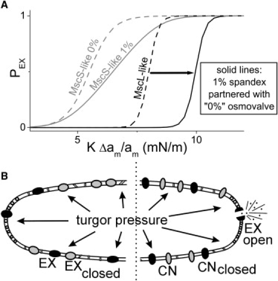
Multiconformation membrane proteins are mechanosensitive (MS) if their conformations displace different bilayer areas. Might MS closed-closed transitions serve as tension buffers, that is, as membrane "spandex"? While bilayer expansion is effectively instantaneous, transitions of bilayer-embedded MS proteins are stochastic (thermally activated) so spandex kinetics would be critical. Here we model generic two-state (contracted/expanded) stochastic spandexes inspired by known bacterial osmovalves (MscL, MscS) then suggest experimental approaches to test for spandex-like behaviors in these proteins. Modeling shows: 1), spandex kinetics depend on the transition state location along an area reaction coordinate; 2), increasing membrane concentration of a spandex right-shifts its midpoint (= tension-Boltzmann); 3), spandexes with midpoints below the activating tension of an osmovalve could optimize osmovalve deployment (required: large midpoint, barrier near the expanded state); 4), spandexes could damp bilayer tension excursions (required: midpoint at target tension, and for speed, barrier halfway between the contracted and expanded states; the larger the spandex Delta-area, the more precise the maintenance of target tension; higher spandex concentrations damp larger amplitude strain fluctuations). One spandex species could not excel as both first line of defense for osmovalve partners and tension damper. Possible interactions among MS closed-closed and closed-open transitions are discussed for MscS- and MscL-like proteins.


Figures

Figure 1
Figure 1
(A) Pure lipid bilayers (top), or ones with nonexpansible proteins (middle), rupture (i.e., lyse) under strain ∼4%, whereas expanding proteins (bottom) could prevent lysis by relief of tension. (B) The spandex model states (contracted, CN; expanded, EX). For a given strain, bilayer tension (= γ) is higher with CN than with EX. (C) Spandex energy landscapes (reaction coordinate: protein area). (Top) A landscape for γ < γ0.5, the spandex midpoint tension, illustrates the model's parameters: ACN, A, and AEX are the areas associated with the CN, transition (barrier) and EX states. With CN taken as the reference state, E and EEX are the energies of the barrier state and of EX. kEX and kCN are the transition rates. (Bottom) As bilayer tension increases, the energy of the EX and barrier states decrease linearly, relative to CN. At γ = γ0.5 CN and EX have the same energy and the probabilities of being expanded and contracted (PEX, PCN) are equal. (D) Three spandex proteins with different values for A (the transition energy maximum's position) and with ACN and AEX fixed at values reported for MscL (17).
Figure 2
Figure 2
(A) PEX(γ) (i.e., tension-Boltzmanns) for three different ΔA spandexes with the same midpoint at 8 mN/m (i.e., slightly lower than that for MscL). ΔA = 0.75 nm2 could be any generic small ΔA protein; ΔA = 1.5 nm2 is in the range calculated for KvAP activation (38); and ΔA = 20.4 nm2 corresponds to MscL (17). (B) A mildly stressed membrane with contracted spandex proteins (i) that is suddenly exposed to a large strain (due, say, to high turgor) (ii) will experience elevated bilayer tension (the spring length, γ, increases) that progressively falls as more spandex proteins expand (iii). If the membrane contained exclusively nonexpansible proteins (iv) it would be more likely to rupture (v). (C) The effect of spandex concentration, Cprot, on PEX (i), and on γ (ii), as a function of imposed strain (multiplied by bilayer compressibility: KΔam/am). (i) Shows the PEX midpoint right-shifting and (ii) shows bilayer tension flattening with increasing Cprot.
Figure 3
Figure 3
For an isolated spandex protein (Cprot = 0), varying barrier location, A, changes the tension-dependence of τprot (the protein time constant, from Eq. 7) (solid lines) without changing the tension-dependence of PEX (from Eq. 3) (dashed lines).
Figure 4
Figure 4
(A) Spandexes with different A values but otherwise identical parameters (γ0.5, ΔA, τmax, Cprot, etc.) relieve tension with different speeds. The time course of bilayer tension after a step strain at t = 0 (0–4.26%) (KΔam/am = 0–10 mN/m) is plotted for three different A spandexes (Cprot = 2%); though all three have the same τmax, they have different τ(γ) behavior (Fig. 3). (B) Two different spandex proteins serve to damp bilayer stress (top) under oscillatory strain (bottom). Damping quality depends on the spandex ΔA. Cprot is varied to keep the product ΔACprot constant: ΔA = 20.4 nm2 and Cprot = 0.02 (solid line) and ΔA = 4.08 nm2 and Cprot = 0.1 (dashed line). The strain is initially set to γ0.5pop = 11.5 mN/m, then oscillates with an amplitude of 1% (corresponding to 2.35 mN/m), with a period of T = 50 ms (i.e., 10 × τmax for these spandexes).
Figure 5
Figure 5
Damped tension excursions. (A) For a quasistatic strain oscillation, the amplitude of the tension variation, Δγ, for three different spandexes varies Cprot. (B) Δγ varies with the period (T) of the oscillating strain (normalized to τmax). For rapid oscillations (toward the left), tension damping is ineffective and the tension oscillation amplitude is that of the strain oscillation (multiplied by compressibility = 2.35 mN/m). For slow strain oscillations (toward the right), the effectiveness varies with Cprot and ΔA. In panels A and B, strain amplitude is 1% (corresponding to 2.35 mN/m). Plots were obtained by solving the model for different Cprot; ΔA and T and are not analytic functions.
Figure 6
Figure 6
Spandex-osmovalve partnerships. (A) For a MscS-like spandex and a MscL-like osmovalve, dotted lines show isolated protein (Cprot = 0 Boltzmanns). If the membrane concentration of MscS-like spandex is increased to 1% (gray solid), this right-shifts (arrow) the Boltzmann of the single MscL-like osmovalve (black solid). (B, left) differential modulation of a MscL population to yield a spandex/osmovalve duo. Anionic lipids positively influence stress-induced opening of MscL (53) and cardiolipin, an anionic negative curvature lipid, segregates to the poles of cylindrical bacteria (54). MscL is ubiquitously dispersed in bacterial membranes (55) (unlike pole-preferring osmotransporter, ProP (56)). If the polar chemical environment is needed for osmovalve opening and not for the preopening expansion, then MscL along the cylindrical surface of the bacterium could exclusively operate in spandex mode. Moreover, since Laplace's law dictates that tension in the cylinder would exceed that at hemispheres, spandex would respond before the polar osmovalves, thus preventing its unnecessary deployment (i.e., effectively right-shifting the MscL-osmovalve, as in panel A). Two spandexes are shown black for the modulated MscL and gray for different protein species to indicate that multiple spandex species (e.g., MscS-like proteins) could participate. (BRight) If proteins in the cylindrical region were unable to expand, the full impact of increasing turgor pressure would be borne by one, or a few, polar osmovalve(s) that would expand, open, and dump osmolytes.

References

    1. Hamill O.P. Twenty odd years of stretch-sensitive channels. Pflügers Arch. Eur. J. Phys. 2006;453:333–351. - PubMed
    1. Sukharev S.I., Anishkin A. Mechanosensitive channels: what can we learn from “simple” model systems? Trends Neurosci. 2004;27:345–351. - PubMed
    1. Booth I.R., Edwards M.D., Black S., Schumann U., Miller S. Mechanosensitive channels in bacteria: signs of closure? Nat. Rev. Microbiol. 2007;5:431–440. - PubMed
    1. Corry B., Martinac B. Bacterial mechanosensitive channels: experiment and theory. Biochim. Biophys. Acta Biomembr. 2008;1778:1859–1870. - PubMed
    1. Evans E., Heinrich V., Ludwig F., Rawicz W. Dynamic tension spectroscopy and strength of biomembranes. Biophys. J. 2003;85:2342–2350. - PMC - PubMed

Publication types

LinkOut - more resources