. 2009 Nov 13;36(3):417-30.
doi: 10.1016/j.molcel.2009.10.012.

Rhythmic PER abundance defines a critical nodal point for negative feedback within the circadian clock mechanism

Affiliations

Rhythmic PER abundance defines a critical nodal point for negative feedback within the circadian clock mechanism

Rongmin Chen et al. Mol Cell. .

Abstract

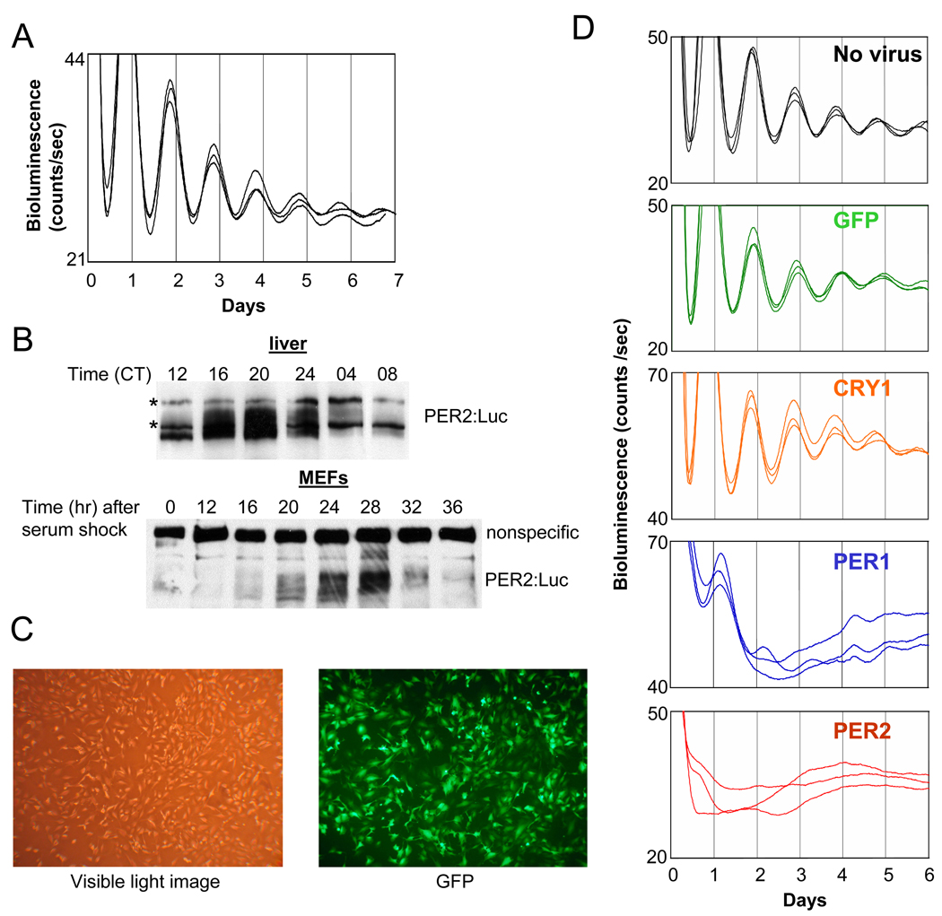
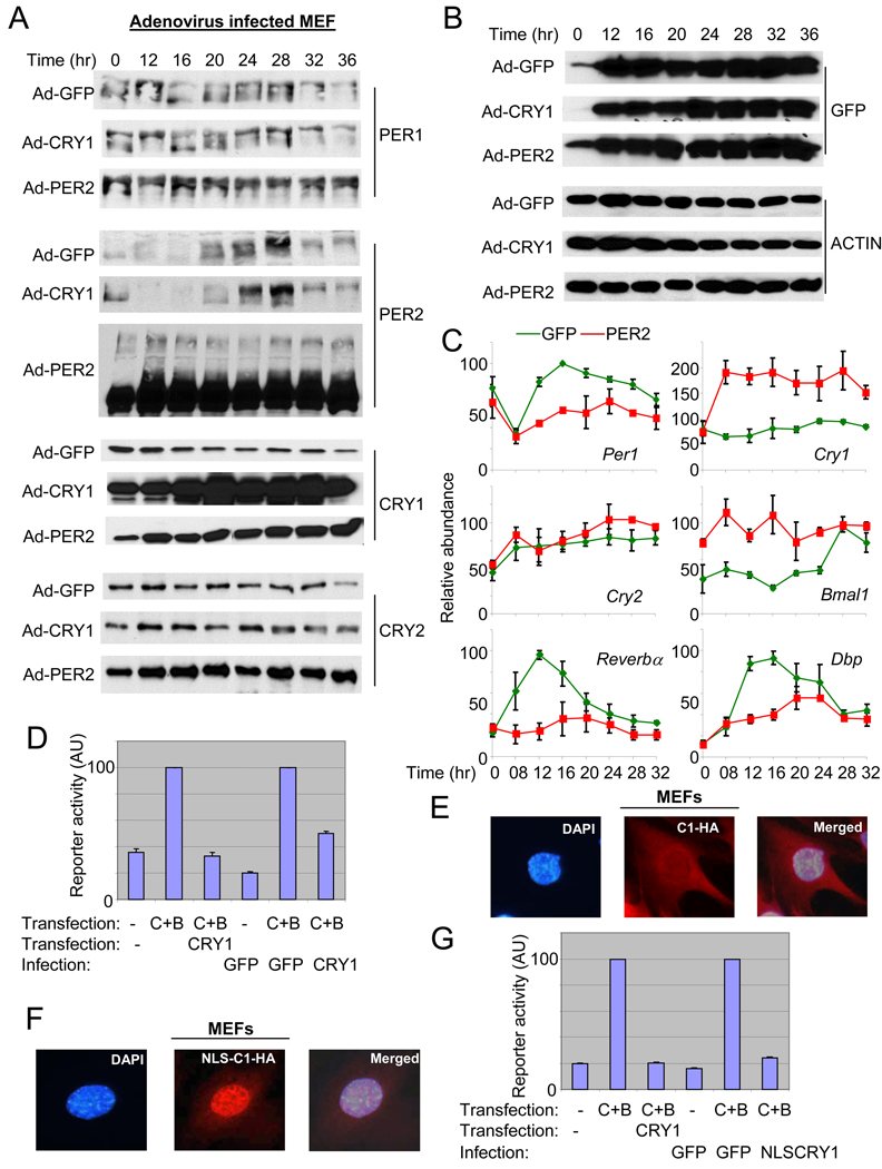
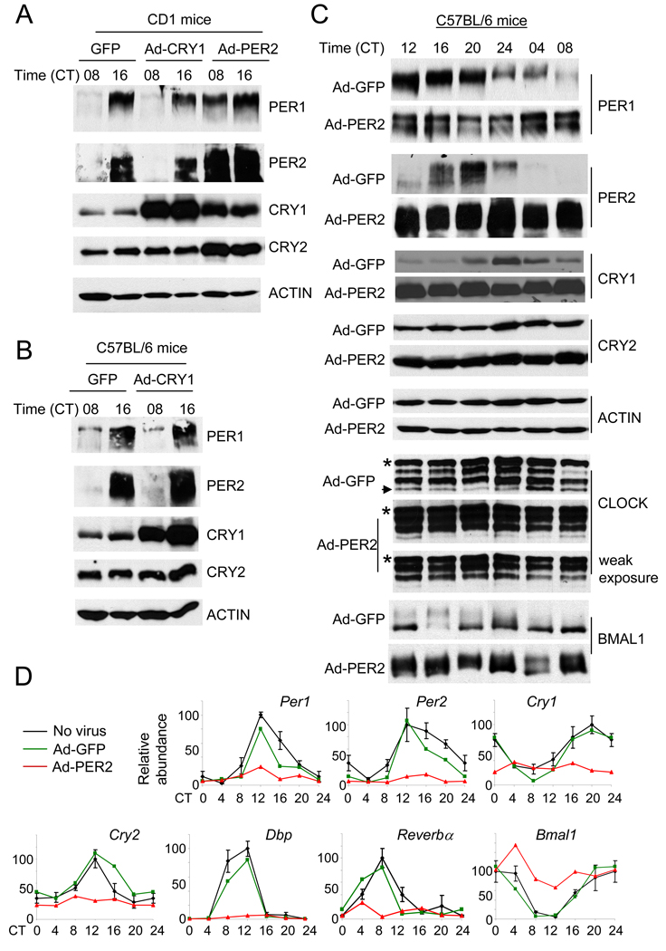
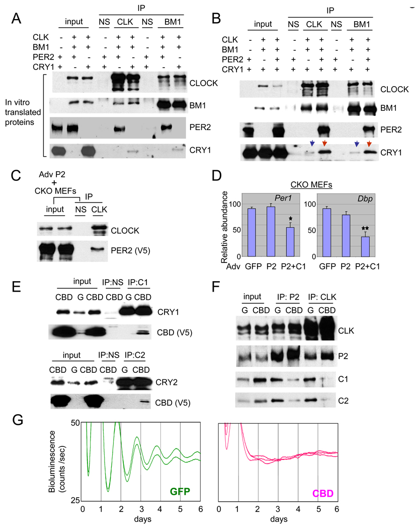
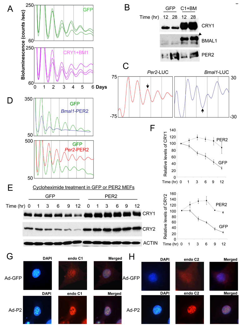
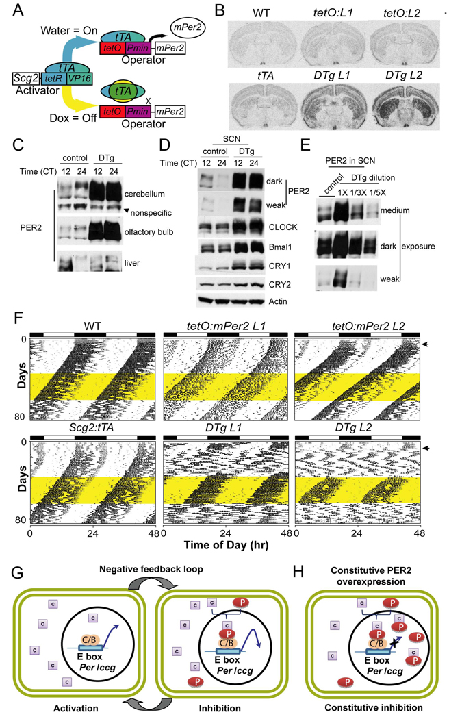
Circadian rhythms in mammals are generated by a transcriptional negative feedback loop that is driven primarily by oscillations of PER and CRY, which inhibit their own transcriptional activators, CLOCK and BMAL1. Current models posit that CRY is the dominant repressor, while PER may play an accessory role. In this study, however, constitutive expression of PER, and not CRY1, severely disrupted the clock in fibroblasts and liver. Furthermore, constitutive expression of PER2 in the brain and SCN of transgenic mice caused a complete loss of behavioral circadian rhythms in a conditional and reversible manner. These results demonstrate that rhythmic levels of PER2, rather than CRY1, are critical for circadian oscillations in cells and in the intact organism. Our biochemical evidence supports an elegant mechanism for the disparity: PER2 directly and rhythmically binds to CLOCK:BMAL1, while CRY only interacts indirectly; PER2 bridges CRY and CLOCK:BMAL1 to drive the circadian negative feedback loop.


Figures

Fig. 1
Fig. 1
Bioluminescence rhythms in MEFs are abolished by constitutively expressed PER, but not by GFP or CRY1. (A) Per2Luc MEFs exhibit circadian bioluminescence rhythms over 7 days after serum shock. (B) PER2:Luciferase fusion protein shows circadian oscillations in abundance and phosphorylation in both liver and MEFs. * indicates nonspecific bands. (C) The adenoviral vector efficiently infects MEFs. MEFs were infected with GFP-adenovirus and fixed 24 hr later to take a visible light image and a fluorescence (GFP) image. (D) MEFs were infected with adenoviral vector expressing GFP, CRY1, PER1 or PER2 or mock infected with medium only (no virus) for 2 hours and then serum shocked for 2 hours before measurement of bioluminescence.
Fig. 2
Fig. 2
Constitutive expression of PER2, and not CRY1, disrupts PER protein rhythms and mRNA rhythms of clock and clock-controlled genes. (A) (B) MEFs were infected and serum shocked as described in Fig 1. Both endogenous (the fusion protein; top band) and exogenous (bottom band) PER2 are shown for PER2 immunoblot of PER2-MEFs. The adenoviral vector has dual CMV promoters, one default CMV promoter for GFP and the other one for the gene of interest (He et al., 1998), in our case PER2 or CRY1. Thus, similar intensities of GFP signal indicate similar viral titers among the three groups. (C) Quantitation of mRNA levels of clock and clock-controlled genes in Adenovirus GFP (green)- and Adenovirus PER2 (red)-MEFs. mRNA levels were measured by quantitative real time PCR. Data are shown as mean+/−SEM of three experiments. For some data points, error bars (if less than 3) are obscured by the square symbols. (D) The viral CRY1 can inhibit CLOCK:BMAL1-driven transcription in reporter assays. The inhibitory activity between plasmid (pcDNA3.1) and adenoviral CRY1 on CLOCK:BMAL1-driven transcription was compared in transient transfection assays. The first three reactions (bars) were done only with plasmid transfection but the other three reactions were performed with transfection (CLOCK and BMAL1) and viral infection (CRY). Results are shown as mean+/−SEM of triplicate samples and are representative of three independent experiments. (E) (F) The viral CRY1-HA was both cytoplasmic and nuclear (E), but the viral NLS-CRY1-HA was predominantly nuclear (F). The overexpressed viral CRY1-HA was visualized with immunofluorescence using anti-HA antibody. These single cells correspond to the cells in white boxes in Suppl Fig 2B–C, magnified as representatives. (G) The inhibitory activity of plasmid (pcDNA3.1)-expressed CRY and adenoviral NLS-CRY1 on CLOCK:BMAL1-driven transcription was compared in COS7 cells, as in (D).
Fig. 3
Fig. 3
Constitutive expression of GFP, CRY1 or PER2 in mouse liver produced similar results as in MEFs. (A) (B) (C) CD-1 (A) and C57BL/6 (B and C) strains were injected with adenovirus expressing GFP, CRY1 or PER2 and sacrificed at the indicated circadian times. Liver extracts were subjected to immunoblotting. Results are representative of at least 2 mice per time point. * indicates a nonspecific band in the CLOCK immunoblot. The arrow indicates a hypophosphorylated isoform, which is much weaker in PER2-liver. Note that the top isoform is much more enriched in the PER2-liver. (D) Quantification of mRNA levels of clock and clock-controlled genes in control (no virus, black), GFP (green)-, and PER2 (red)-liver. mRNA levels were measured by quantitative real-time PCR. Results are average+/−SEM of three mice for controls, and representative of two mice per time point for GFP and PER2-adenovirus injected mice. The data for the second set of the GFP and PER2 mice are shown in Suppl Fig 6. Levels were normalized so that the highest levels of the control mice equaled 100 arbitrary units. The CT 0 data were plotted twice as CT 0 and 24.
Fig. 4
Fig. 4
In GFP- or CRY-overexpressing liver, rhythmic interaction between CLOCK:BMAL1 and PER/CRY is maintained, but the interaction is constitutively elevated in PER2-overexpressing liver and almost absent in Per1/2ldc mutant mice. (A) Liver samples at CT 08 and 16 in Fig 4A (CD1 mice) were subjected to IP for CLOCK and BMAL1 and the resulting immunocomplexes were immunoblotted for clock proteins. Two arrow heads indicate two hyperphosphorylated isoforms of CLOCK compared to the other two isoforms as has been shown previously (Lee et al., 2001). The asterisk denotes a nonspecific band in the BMAL1 immunoblot ensuring equal loading of total protein. (B) IP for CLOCK and BMAL1 was performed using liver samples at CT06 and 18 from Per1/2ldc mutant and matching wt mice. To compensate for lower amounts of clock proteins in the mutant mice, twice as much starting material was used for the IP experiments. Note that CLOCK is hypophosphorylated, but BMAL1 is hyperphosphorylated in the mutant mice.
Fig. 5
Fig. 5
PER is the scaffolding molecule between the activator complex (CLOCK:BMAL1) and the negative complex (PER:CRY). (A) PER2 has a much higher affinity than CRY1 for binding to CLOCK:BMAL1. Equal moles of in vitro translated proteins were mixed in different combinations and subjected to IP for CLOCK or BMAL1. (B) CRY1 copurified with CLOCK and BMAL1 is dramatically increased in the presence of PER2 in vitro. Equimolar amounts of the in vitro translated proteins were mixed as indicated and subjected to IP for CLOCK or BMAL1. Note that copurified CRY1 levels are dramatically different when PER2 is absent (the blue arrows) and present (the red arrows). (C) PER2 does not require CRY for binding CLOCK. PER2-V5 adenovirus was infected into primary Cry double-mutant MEFs to test if PER2 can interact with CLOCK:BMAL1 in the absence of CRYs. (D) Adenoviral overexpression of PER2 in the Cry double-knockout (CKO) MEFs does not suppress endogenous mRNA expression of Per1 and Dbp, but co-overexpression of PER2 and CRY1 does. Average+/−SEM of three experiments is shown. GFP and PER2+CRY1 samples differed significantly (Student t-test; one star: p<0.05; two stars: p<0.01). (E) CRY-binding domain of PER2 (CBD) interacts with endogenous CRY1 and 2. MEFs were infected with GFP or CBD (CBD-V5) adenovirus and subjected to IP for CRY1 or CRY2. (F) CBD disrupts CRY interaction with clock protein complexes. Results from IP for BMAL1 are shown in Suppl. Fig 11. (G) PER2:CRY interaction is essential for circadian rhythm generation. When the interaction is disrupted by CBD, overt rhythms were completely abolished.
Fig. 6
Fig. 6
Per oscillation plays a dominant role in circadian rhythm generation. (A) Overexpression of both CRY1 and BMAL1 does not affect the circadian rhythms. MEFs were co-infected with CRY1 and BMAL1 adenoviruses. (B) PER2 remains rhythmic in MEFs overexpressing CRY1 and BMAL1. MEFs were harvested 12 and 28 hr after GFP or CRY1/BMAL1 adenovirus infection and serum shock. The arrow head indicates the exogenous BMAL1 (2XHA-BMAL1). (C) Per2 and Bmal1 promoters drive antiphasic oscillations in MEFs. Adenovirus expressing Luciferase under Per2 or Bmal1 promoter was infected into wt MEFs and bioluminescence rhythms were measured. The arrows indicate the antiphasic oscillations by two promoters. (D) PER2 expression from the Bmal1 promoter severely compromised the circadian rhythms in Per2Luc MEFs, whereas that from Per2 promoter did not disrupt the circadian rhythms. (E) (F) Extended half-lives of endogenous CRYs in PER2-MEFs. To measure half-lives of endogenous CRYs, cycloheximide was added 12 hrs after GFP or PER2 adenovirus infection and serum shock. (F) shows mean +/−SEM of three experiments. (G) (H) Nuclear staining of endogenous CRYs is more pronounced in PER2-MEFs than in GFP-MEFs.
Fig. 7
Fig. 7
PER2 overexpression in the brain, including SCN, abolished locomotor activity rhythms in constant darkness (DD). (A) Conditional Per2 expression. The system works by driving the tetracycline transactivator (tTA) expression from a tissue-specific promoter (Scg2), which binds the tetracycline operator (tetO) and drives the transcription of Per2 through a minimal promoter sequence (Pmin). However, doxycycline (Dox) potently inhibits tTA, turning off Per2 transgene expression. (B) In situ hybridization for Per2 confirms exogenous expression throughout the brain with enriched expression in the SCN. Representative sections encompassing the SCN are shown for six genotypes: wild type (WT); tetO:Per2 Line1 Only (tetO:L1); tetO:Per2 Line 2 Only (tetO:L2); Scg2:tTA Only (tTA); tetO:Per2 Line 1/Scg2:tTA (DTg L1); tetO:Per2 Line 2/Scg2:tTA (DTg L2). Levels of Per2 mRNA expression in the SCN of double transgenic (DTg) mice were 2–3 fold higher than those in the SCN of WT and other control mice (Suppl. Fig 14A). (C) (D) PER2 levels in DTg were higher in olfactory bulbs (OB), cerebellum and SCN than those in control mice. The single transgenic (tetO:L2) mouse was used as the control. The tissues from control and DTg mice were collected at CT12 and 24. Unlike OB and cerebellum, there was no sign of overexpression in liver. Note that the phase of PER2 rhythm in control mice is different between SCN and the other brain areas. (E) The serial dilution samples of the DTg SCN extract at CT12 indicate that the levels of PER2 overexpression were 3–5 fold higher than those of PER2 in the control mice at CT12. Quantitative chemiluminescence imaging is shown in Suppl Fig 15. (F) Disrupted circadian rhythms of PER2-overexpressing double-transgenic mice. Representative actograms for the two double transgenic lines show a loss of rhythmicity in DD when compared to WT and single transgenic controls. Locomotor behavior returns to a WT state in the double transgenic animals with a treatment of 10μg/ml dox. This treatment can then be reversed with a water washout converting the double transgenic animals back to a state with no circadian rhythmicity. The yellow shading denotes the time of dox administration in DD. (G) A revised mammalian circadian clock model. When PER is absent, CRY is predominantly cytoplasmic and transcriptional activation by CLOCK:BMAL1 is permitted (activation phase). However, when PER abundance reaches a certain level, PER:CRY complexes enter or accumulate in the nucleus. In the nucleus, PER directly binds the CLOCK:BMAL1 complex to mediate the negative feedback effects of the PER:CRY complex (inhibition phase of the negative feedback loop). (H) In the presence of constitutively overexpressed PER, excess PER:CRY complexes accumulate in the nucleus and constitutively inhibit CLOCK:BMAL1 complex, leading to constitutive inhibition of the negative feedback loop.

References

    1. Allada R, Emery P, Takahashi JS, Rosbash M. Stopping time: the genetics of fly and mouse circadian clocks. Annu Rev Neurosci. 2001;24:1091–1119. - PubMed
    1. Aronson BD, Johnson KA, Loros JJ, Dunlap JC. Negative feedback defining a circadian clock: autoregulation of the clock gene frequency. Science. 1994;263:1578–1584. - PubMed
    1. Bae K, Jin X, Maywood ES, Hastings MH, Reppert SM, Weaver DR. Differential functions of mPer1, mPer2, and mPer3 in the SCN circadian clock. Neuron. 2001;30:525–536. - PubMed
    1. Baylies MK, Bargiello TA, Jackson FR, Young MW. Changes in abundance or structure of the per gene product can alter periodicity of the Drosophila clock. Nature. 1987;326:390–392. - PubMed
    1. Bunger MK, Wilsbacher LD, Moran SM, Clendenin C, Radcliffe LA, Hogenesch JB, Simon MC, Takahashi JS, Bradfield CA. Mop3 is an essential component of the master circadian pacemaker in mammals. Cell. 2000;103:1009–1017. - PMC - PubMed

Publication types

MeSH terms