E6 proteins from diverse papillomaviruses self-associate both in vitro and in vivo
- PMID: 19917295
- PMCID: PMC3900769
- DOI: 10.1016/j.jmb.2009.11.022
E6 proteins from diverse papillomaviruses self-associate both in vitro and in vivo
Abstract
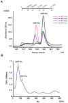
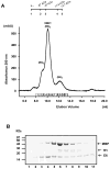
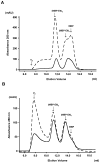
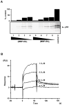
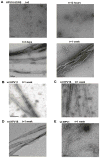
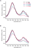
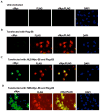
Papillomavirus E6 oncoproteins bind and often provoke the degradation of many cellular proteins important for the control of cell proliferation and/or cell death. Structural studies on E6 proteins have long been hindered by the difficulties of obtaining highly concentrated samples of recombinant E6. Here, we show that recombinant E6 proteins from eight human papillomavirus strains and one bovine papillomavirus strain exist as oligomeric and multimeric species. These species were characterized using a variety of biochemical and biophysical techniques, including analytical gel filtration, activity assays, surface plasmon resonance, electron microscopy and Fourier transform infrared spectroscopy. The characterization of E6 oligomers is facilitated by the fusion to the maltose binding protein, which slows the formation of higher-order multimeric species. The proportion of each oligomeric form varies depending on the viral strain considered. Oligomers appear to consist of folded units, which, in the case of high-risk mucosal human papillomavirus E6, retain binding to the ubiquitin ligase E6-associated protein and the capacity to degrade the proapoptotic protein p53. In addition to the small-size oligomers, E6 proteins spontaneously assemble into large organized multimeric structures, a process that is accompanied by a significant increase in the beta-sheet secondary structure content. Finally, co-localisation experiments using E6 equipped with different tags further demonstrate the occurrence of E6 self-association in eukaryotic cells. The ensemble of these data suggests that self-association is a general property of E6 proteins that occurs both in vitro and in vivo and might therefore be functionally relevant.
Copyright 2009 Elsevier Ltd. All rights reserved.
Figures



Similar articles
-
Strategies for bacterial expression of protein-peptide complexes: application to solubilization of papillomavirus E6.Protein Expr Purif. 2011 Nov;80(1):8-16. doi: 10.1016/j.pep.2011.06.013. Epub 2011 Jul 14. Protein Expr Purif. 2011. PMID: 21777678 Free PMC article.
-
Determinants of stability for the E6 protein of papillomavirus type 16.J Mol Biol. 2009 Mar 6;386(4):1123-37. doi: 10.1016/j.jmb.2009.01.018. J Mol Biol. 2009. PMID: 19244625 Free PMC article.
-
Purification and biophysical characterization of a minimal functional domain and of an N-terminal Zn2+-binding fragment from the human papillomavirus type 16 E6 protein.Biochemistry. 2001 Feb 6;40(5):1196-204. doi: 10.1021/bi001837+. Biochemistry. 2001. PMID: 11170444
-
Formation of well-defined soluble aggregates upon fusion to MBP is a generic property of E6 proteins from various human papillomavirus species.Protein Expr Purif. 2007 Jan;51(1):59-70. doi: 10.1016/j.pep.2006.07.029. Epub 2006 Sep 9. Protein Expr Purif. 2007. PMID: 17055740
-
Predicted alpha-helix/beta-sheet secondary structures for the zinc-binding motifs of human papillomavirus E7 and E6 proteins by consensus prediction averaging and spectroscopic studies of E7.Biochem J. 1996 Oct 1;319 ( Pt 1)(Pt 1):229-39. doi: 10.1042/bj3190229. Biochem J. 1996. PMID: 8870673 Free PMC article.
Cited by
-
Parallel β-sheet vibrational couplings revealed by 2D IR spectroscopy of an isotopically labeled macrocycle: quantitative benchmark for the interpretation of amyloid and protein infrared spectra.J Am Chem Soc. 2012 Nov 21;134(46):19118-28. doi: 10.1021/ja3074962. Epub 2012 Nov 9. J Am Chem Soc. 2012. PMID: 23113791 Free PMC article.
-
Strategies for bacterial expression of protein-peptide complexes: application to solubilization of papillomavirus E6.Protein Expr Purif. 2011 Nov;80(1):8-16. doi: 10.1016/j.pep.2011.06.013. Epub 2011 Jul 14. Protein Expr Purif. 2011. PMID: 21777678 Free PMC article.
-
Structural basis for hijacking of cellular LxxLL motifs by papillomavirus E6 oncoproteins.Science. 2013 Feb 8;339(6120):694-8. doi: 10.1126/science.1229934. Science. 2013. PMID: 23393263 Free PMC article.
-
Discrimination of Human Cell Lines by Infrared Spectroscopy and Mathematical Modeling.Iran J Pharm Res. 2015 Summer;14(3):803-10. Iran J Pharm Res. 2015. PMID: 26330868 Free PMC article.
-
Patterns Prediction of Chemotherapy Sensitivity in Cancer Cell lines Using FTIR Spectrum, Neural Network and Principal Components Analysis.Iran J Pharm Res. 2012 Spring;11(2):401-10. Iran J Pharm Res. 2012. PMID: 24250464 Free PMC article.
References
-
- zur Hausen H. Papillomaviruses in human cancers. Proc Assoc Am Physicians. 1999;111:581–587. - PubMed
-
- Bosch FX, Manos MM, Munoz N, Sherman M, Jansen AM, Peto J, Schiffman MH, Moreno V, Kurman R, Shah KV. Prevalence of human papillomavirus in cervical cancer: a worldwide perspective. International biological study on cervical cancer (IBSCC) Study Group. J Natl Cancer Inst. 1995;87:796–802. - PubMed
-
- Pfister H. Chapter 8: Human papillomavirus and skin cancer. J Natl Cancer Inst Monogr. 2003;31:52–56. - PubMed
-
- Lambert PF, Baker CC, Howley PM. The genetics of bovine papillomavirus type 1. Annu Rev Genet. 1988;22:235–58. - PubMed
Publication types
MeSH terms
Substances
Grants and funding
LinkOut - more resources
Full Text Sources
Other Literature Sources
Research Materials
Miscellaneous

