

Mechanisms of tissue injury in renal artery stenosis: ischemia and beyond
- PMID: 19917330
- PMCID: PMC2800096
- DOI: 10.1016/j.pcad.2009.09.002
Mechanisms of tissue injury in renal artery stenosis: ischemia and beyond
Abstract
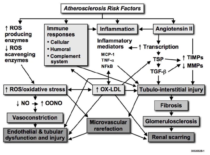
Renal injury distal to an atherosclerotic renovascular obstruction reflects multiple intrinsic factors producing parenchymal tissue injury. Atherosclerotic disease pathways superimposed on renal arterial obstruction may aggravate damage to the kidney and other target organs, and some of the factors activated by renal artery stenosis may in turn accelerate the progression of atherosclerosis. This cross-talk is mediated through amplified activation of renin-angiotensin system, oxidative stress, inflammation, and fibrosis-pathways notoriously involved in renal disease progression. Oxidation of lipids also accelerates the development of fibrosis in the stenotic kidney by amplifying profibrotic mechanisms and disrupting tissue remodeling. The extent to which actual ischemia modulates injury in the stenotic kidney has been controversial, partly because the decrease in renal oxygen consumption usually parallels a decrease in renal blood flow, and because renal vein oxygen pressure in the affected kidney is not decreased. However, recent data using novel methodologies demonstrate that intra-renal oxygenation is heterogeneously affected in different regions of the kidney. Activation of such local injury within the kidney may lead to renal dysfunction and structural injury, and ultimately unfavorable and irreversible renal outcomes. Identification of specific pathways producing progressive renal injury may enable development of targeted interventions to block these pathways and preserve the stenotic kidney.
Figures
References
-
- Hansen KJ, Edwards MS, Craven TE, Cherr GS, Jackson SA, Appel RG, Burke GL, Dean RH. Prevalence of renovascular disease in the elderly: a population-based study. J Vasc Surg. 2002;36:443–51. - PubMed
-
- Uzu T, Takeji M, Yamada N, Fujii T, Yamauchi A, Takishita S, Kimura G. Prevalence and outcome of renal artery stenosis in atherosclerotic patients with renal dysfunction. Hypertens Res. 2002;25:537–42. - PubMed
-
- Vashist A, Heller EN, Brown EJ, Jr, Alhaddad IA. Renal artery stenosis: a cardiovascular perspective. Am Heart J. 2002;143:559–64. - PubMed
-
- Manjunath G, Tighiouart H, Ibrahim H, Macleod B, Salem D, Griffith J, Coresh J, Levey A, Sarnak M. Level of kidney function as a risk factor for atherosclerotic cardiovascular outcomes in the community. J Am Coll Cardiol. 2003;41:47–55. - PubMed
-
- Conlon PJ, Little MA, Pieper K, Mark DB. Severity of renal vascular disease predicts mortality in patients undergoing coronary angiography. Kidney Int. 2001;60:1490–7. - PubMed
Publication types
MeSH terms
Grants and funding
LinkOut - more resources
Full Text Sources
Medical
