. 2009 Nov 19;6(5):409-21.
doi: 10.1016/j.chom.2009.10.004.

Tetherin-driven adaptation of Vpu and Nef function and the evolution of pandemic and nonpandemic HIV-1 strains

Affiliations

Tetherin-driven adaptation of Vpu and Nef function and the evolution of pandemic and nonpandemic HIV-1 strains

Daniel Sauter et al. Cell Host Microbe. .

Abstract

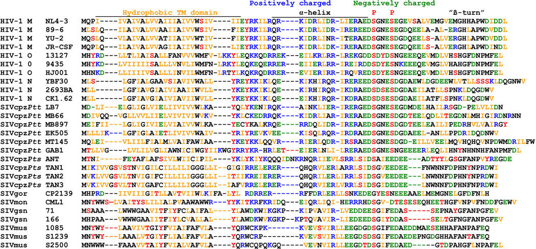
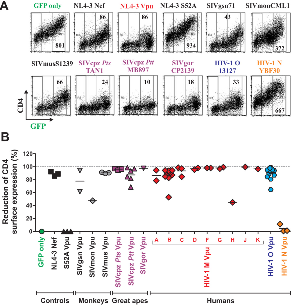
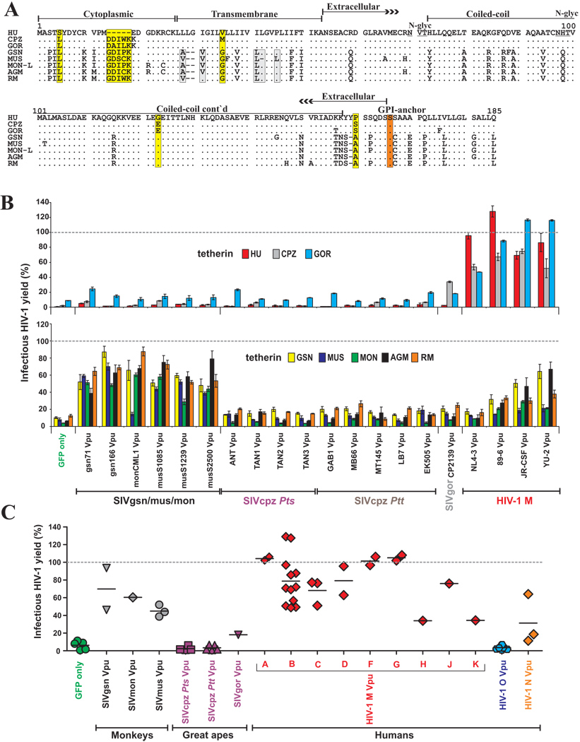
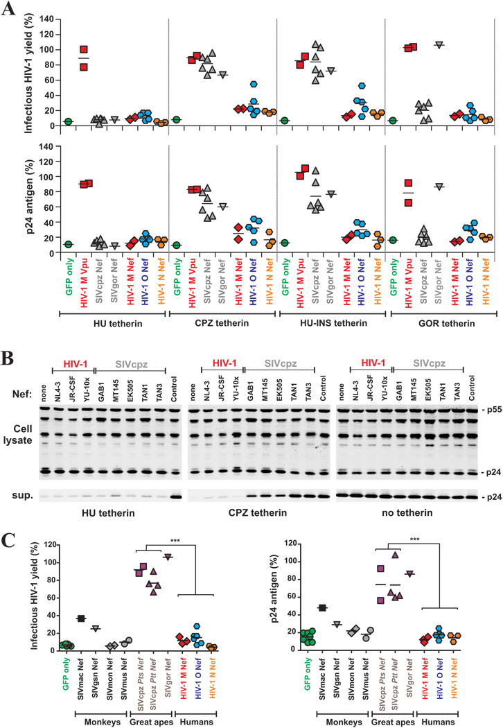
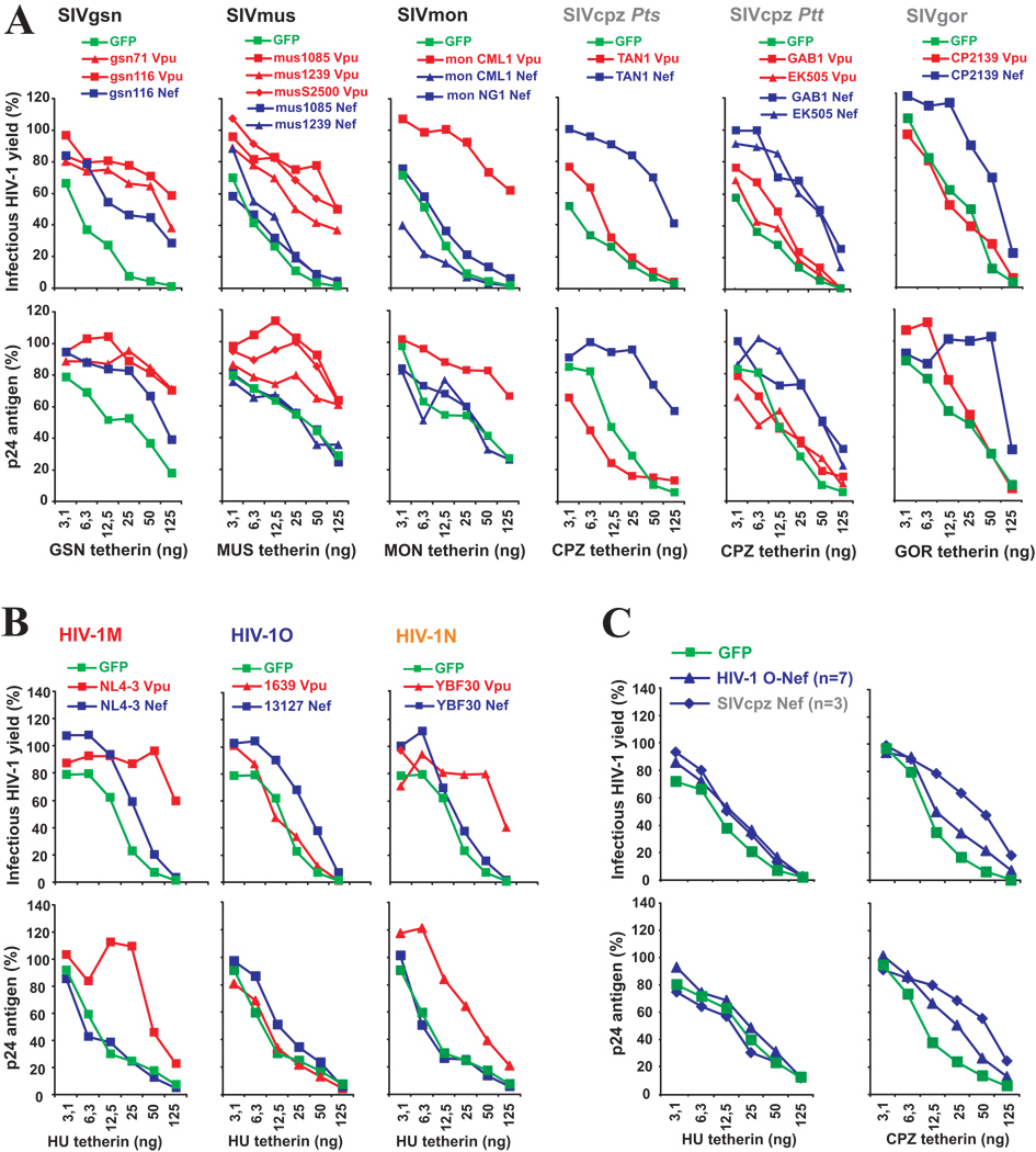
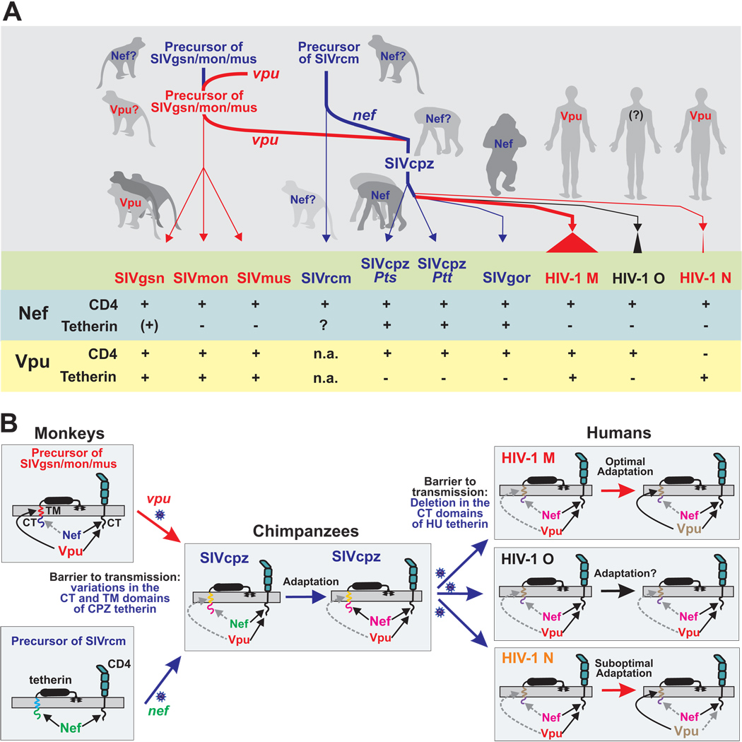
Vpu proteins of pandemic HIV-1 M strains degrade the viral receptor CD4 and antagonize human tetherin to promote viral release and replication. We show that Vpus from SIVgsn, SIVmus, and SIVmon infecting Cercopithecus primate species also degrade CD4 and antagonize tetherin. In contrast, SIVcpz, the immediate precursor of HIV-1, whose Vpu shares a common ancestry with SIVgsn/mus/mon Vpu, uses Nef rather than Vpu to counteract chimpanzee tetherin. Human tetherin, however, is resistant to Nef and thus poses a significant barrier to zoonotic transmission of SIVcpz to humans. Remarkably, Vpus from nonpandemic HIV-1 O strains are poor tetherin antagonists, whereas those from the rare group N viruses do not degrade CD4. Thus, only HIV-1 M evolved a fully functional Vpu following the three independent cross-species transmissions that resulted in HIV-1 groups M, N, and O. This may explain why group M viruses are almost entirely responsible for the global HIV/AIDS pandemic.


Figures

Figure 1
Figure 1
Alignment of HIV-1 and SIV Vpu amino acid sequences. The NL4-3 Vpu sequence is shown on top for comparison. The hydrophobic transmembrane (TM) domain, the central charged region, the position of two serine phosphorylation sites and a β-turn motif in the NL4-3 Vpu protein are indicated. Dashes indicate gaps introduced to optimize the alignment. Acidic residues (E, D) are highlighted in green, basic residues (K, R) in blue, hydrophobic residues (I, V, L) in orange and residues that can potentially be phosphorylated (S, T, Y) in red.
Figure 2
Figure 2. Suppression of CD4 surface expression by HIV-1 and SIV Vpus
(A) FACS analysis of 293T cells cotransfected with a CD4 expression vector and pCGCG plasmids expressing GFP alone (GFP only) or together with the indicated vpu alleles. Constructs expressing the NL4-3 Nef and the mutant NL4-3 S52A Vpu were used as positive and negative controls, respectively. (B) Reduction of Vpu-mediated CD4 expression in 293T cells. Shown is the reduction in the levels of CD4 cell surface expression relative to those measured in cells transfected with the GFP only control vector. The range of GFP expression used for the calculation is indicated in panel A. Each symbol represents one of the 57 vpu alleles examined (Table S1) or the indicated controls. Shown are average values derived from three experiments. The GFP control is color coded green, vpu alleles derived from SIVgsn, SIVmon and SIVmus grey, SIVcpz and SIVgor magenta, HIV-1 M red, O blue and N orange.
Figure 3
Figure 3. Tetherin antagonism by primate lentiviral Vpu proteins
(A) Alignment of tetherin amino acid sequences from humans (HU), chimpanzees (CPZ), gorillas (GOR), Greater spot-nosed monkeys (GSN), Mustached monkeys (MUS), Mona monkeys (MON-L, C. mona lowei), African green monkeys (AGM) and rhesus macaques (RM). Amino acid identity is indicated by dots and gaps by dashes. Differences between HU and CPZ tetherins are highlighted by yellow boxes and variations in the TM domain of monkey tetherins by grey boxes. Known domains, the serine GPI anchor site (red box) and two potential glycosylation sites (underlined), are indicated. (B) Effect of various Vpus on infectious virus release in the presence of the indicated tetherin molecules. 293T cells were cotransfected with HIV-1 ΔVpu NL4-3 (2 µg), pCGCG vectors expressing GFP alone or together with Vpu (500 ng), and tetherin expression constructs (50 ng). Viral supernatants were obtained 2 days later and used to measure the quantity of infectious HIV-1 in the culture supernatants by infecting TZM-bl indicator cells. Shown are average values ±SD (n=3) of infectious virion yield relative to those obtained in the absence of tetherin expression vector (100%). The results were confirmed in at least two independent experiments. (C) Infectious virus release from 293T cells expressing vpu alleles from the indicated primate lentiviruses and tetherins from their respective host species. The experiments were performed as described in panel B. Each symbol represents the average infectious virus release (n=3) obtained in the presence of one of the 55 individual vpu alleles analyzed.
Figure 4
Figure 4. Tetherin antagonism by primate lentiviral Nef proteins
(A) Infectious virus and p24 antigen yield from 293T cells cotransfected with the proviral HIV-1 NL4-3 ΔVpu ΔNef construct (Rucker et al., 2004) containing disrupted vpu and nef genes (2 µg), pCGCG vectors expressing the indicated nef alleles or the NL4-3 Vpu protein (500 ng), in combination with plasmids expressing the HU, CPZ or GOR tetherins or a Hu tetherin variant in which the N-terminal deletion was restored (HU-INS) (50 ng). The upper panel shows the average values derived from triplicate infections of TZM-bl indicator cells and the lower panel the quantity of p24 antigen in the culture supernatant. All values are shown relative to those obtained in the absence of tetherin expression vector (100%) the levels of cellular p24 expression did not differ significantly (data not shown). (B) Western blot analysis of cell and virion lysates following cotransfection of 293T cells with vpu-defective proviral Vpu/Nef/GFP HIV-1 NL4-3 constructs expressing the indicated nef alleles and 50 ng of empty vector (control) or HU or CPZ tetherin-HA expression plasmids. Cell and virion lysates were probed with an anti HIV-1 capsid p24 monoclonal antibody. A proviral HIV-1 NL4-3 construct containing defects in both vpu and nef genes was used in the “none” and “control” lanes. Sup., cell culture supernatant. (C) Infectious virus release from 293T cells expressing nef alleles from the indicated primate lentiviruses and tetherins from their respective host species. The experiments were performed as described in panel A. The symbols represent the average infectious virus release (n=3) obtained in the presence of one of the 25 nef alleles analyzed. ***, p<0.0001.
Figure 5
Figure 5. Titration of anti-tetherin antagonism
(A, B) Virus release from 293T cells following transfection with 2 µg of a ΔVpu/ Nef proviral NL4-3 construct, 500 ng of SIV (A) or HIV-1 (B) Vpu (color coded red) or Nef (color coded blue) expression constructs and varying amounts of plasmids expressing the tetherin molecules from the respective host species. Infectious virus was determined by infection of TZM-bl indicator cells (upper panels) and p24 ELISA (lower panels) is shown as a percentage of that detected in the absence of tetherin (100%). All infections shown in panels A–C were performed in triplicate and the results were confirmed in an independent experiment. (C) HIV-1 O Nefs are inactive against HU tetherin. The assays were performed as described in panels A and B. Shown are average values obtained using seven group O (HJ428, HJ036, HJ736, HJ100, HJ256, 13127 and 8161) and three SIVcpz Ptt (Gab1, MT145, MB897) nef alleles.
Figure 6
Figure 6. Hypothetical model of tetherin driven Vpu and Nef evolution
(A) Schematic of the acquisition of vpu and the subsequent transfer of vpu and nef genes from monkeys to chimpanzees and to humans by zoonotic primate lentiviral transmissions. The events that led to the emergence of pandemic HIV-1 group M are indicated by thick lines. Nef-mediated tetherin antagonism is indicated by blue and Vpu-mediated tetherin antagonism by red lines, respectively. The lower part summarized the results of the present study and previous data on Nef function. Note that the upper part is hypothetical as the ancient vpu and nef genes are not available for analysis. (B) Tetherin-driven evolution of Vpu and Nef function. Nef is present in all primate lentiviruses and interacts with the cytoplasmic tails (CTs) of tetherin and CD4. Vpu was initially acquired by a precursor of SIVgsn/mus/mon and also targets the CT of CD4 but antagonizes tetherin by interacting with its transmembrane (TM) domain. The vpu-containing precursor of SIVgsn/mon/mus (upper left) recombined with that of SIVrcm (lower left) in chimpanzees and the resulting recombinant virus, i.e. SIVcpz, was subsequently transmitted to humans. Since Nef and Vpu inhibit tetherin in a species-specific manner they were most likely initially poorly active against CPZ tetherin. Subsequently, Nef evolved to become an effective tetherin antagonist in chimpanzees (middle). Upon cross-species transmission of SIVcpz to humans, however, this Nef function was most likely disrupted by a unique deletion in the CT of the human restriction factor. Subsequently, Vpu evolved to counteract HU tetherin during the emergence of pandemic HIV-1 M strains (upper right). In comparison, HIV-1 O Vpus remained poor tetherin antagonists (middle right) and those from HIV-1 N lost the CD4 degradation activity (lower right). Matching colors indicate that Vpu and Nef recognize their TM and CT interaction sites in the tetherin molecules. Broken grey arrows indicate disrupted or impaired activities.

Comment in

References

    1. Bailes E, Gao F, Bibollet-Ruche F, Courgnaud V, Peeters M, Marx PA, Hahn BH, Sharp PM. Hybrid origin of SIV in chimpanzees. Science. 2003;300:1713. - PubMed
    1. Barlow KL, Ajao AO, Clewley JP. Characterization of a novel simian immunodeficiency virus (SIVmonNG1) genome sequence from a mona monkey (Cercopithecus mona) J. Virol. 2003;77:6879–6888. - PMC - PubMed
    1. Bour S, Strebel K. The human immunodeficiency virus (HIV) type 2 envelope protein is a functional complement to HIV type 1 Vpu that enhances particle release of heterologous retroviruses. J. Virol. 1996;70:8285–8300. - PMC - PubMed
    1. Bour S, Schubert U, Strebel K. The human immunodeficiency virus type 1 Vpu protein specifically binds to the cytoplasmic domain of CD4: implications for the mechanism of degradation. J. Virol. 1995;69:1510–1520. - PMC - PubMed
    1. Butler IF, Pandream I, Marxm PA, Apetrei C. HIV genetic diversity: biological and public health consequences. Curr. HIV Res. 2007;5:23–45. - PubMed

Publication types

MeSH terms

Substances

Associated data