. 2009 Nov 19;6(5):433-45.
doi: 10.1016/j.chom.2009.09.013.

Cowpox virus inhibits the transporter associated with antigen processing to evade T cell recognition

Affiliations

Cowpox virus inhibits the transporter associated with antigen processing to evade T cell recognition

Dina Alzhanova et al. Cell Host Microbe. .

Abstract

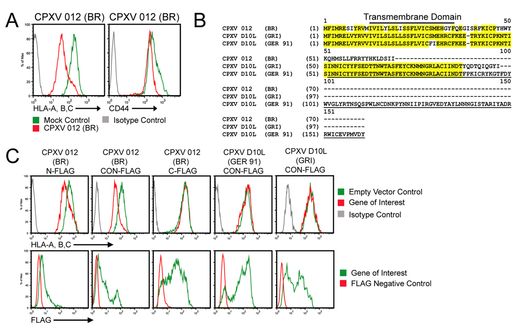
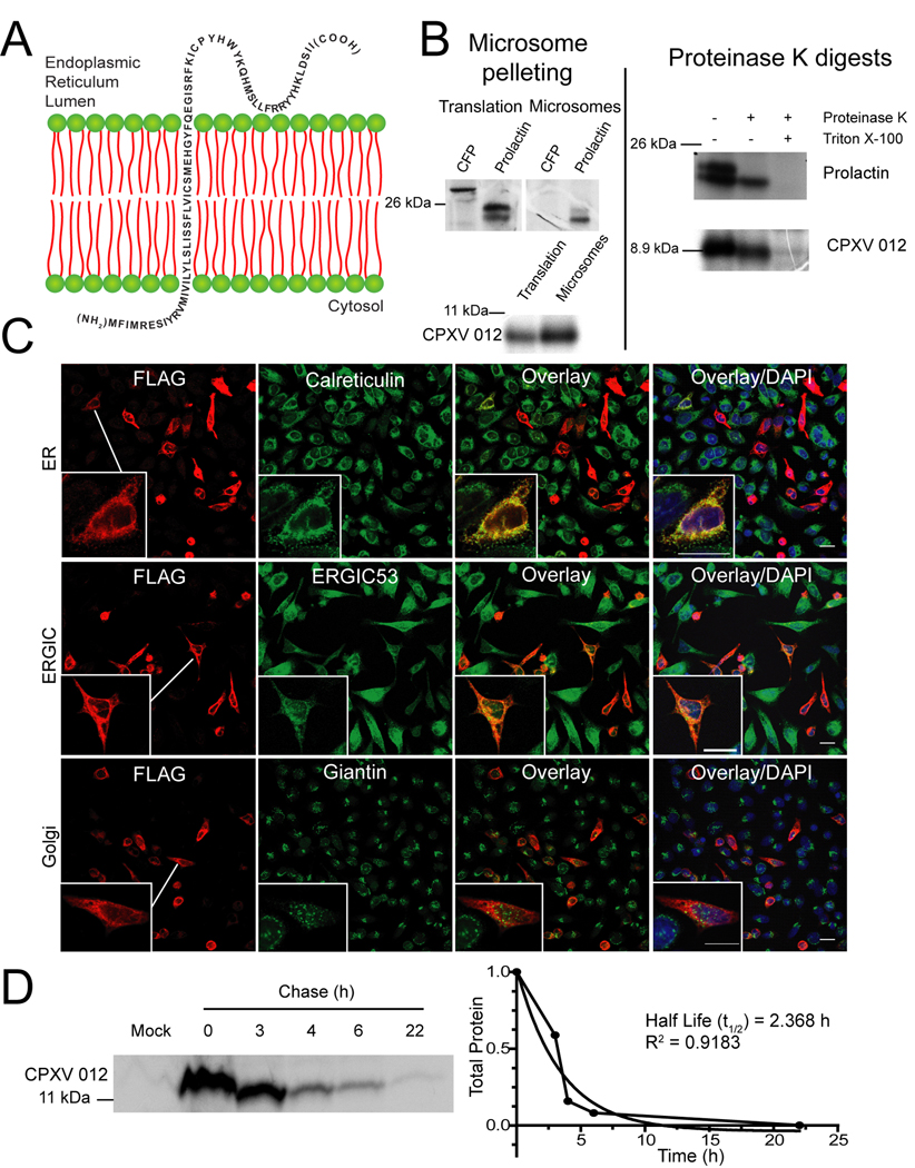
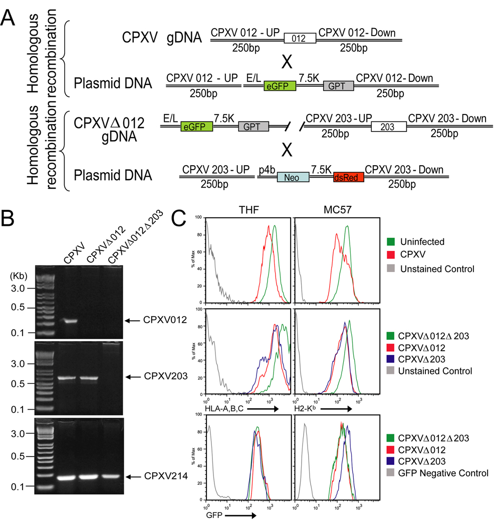
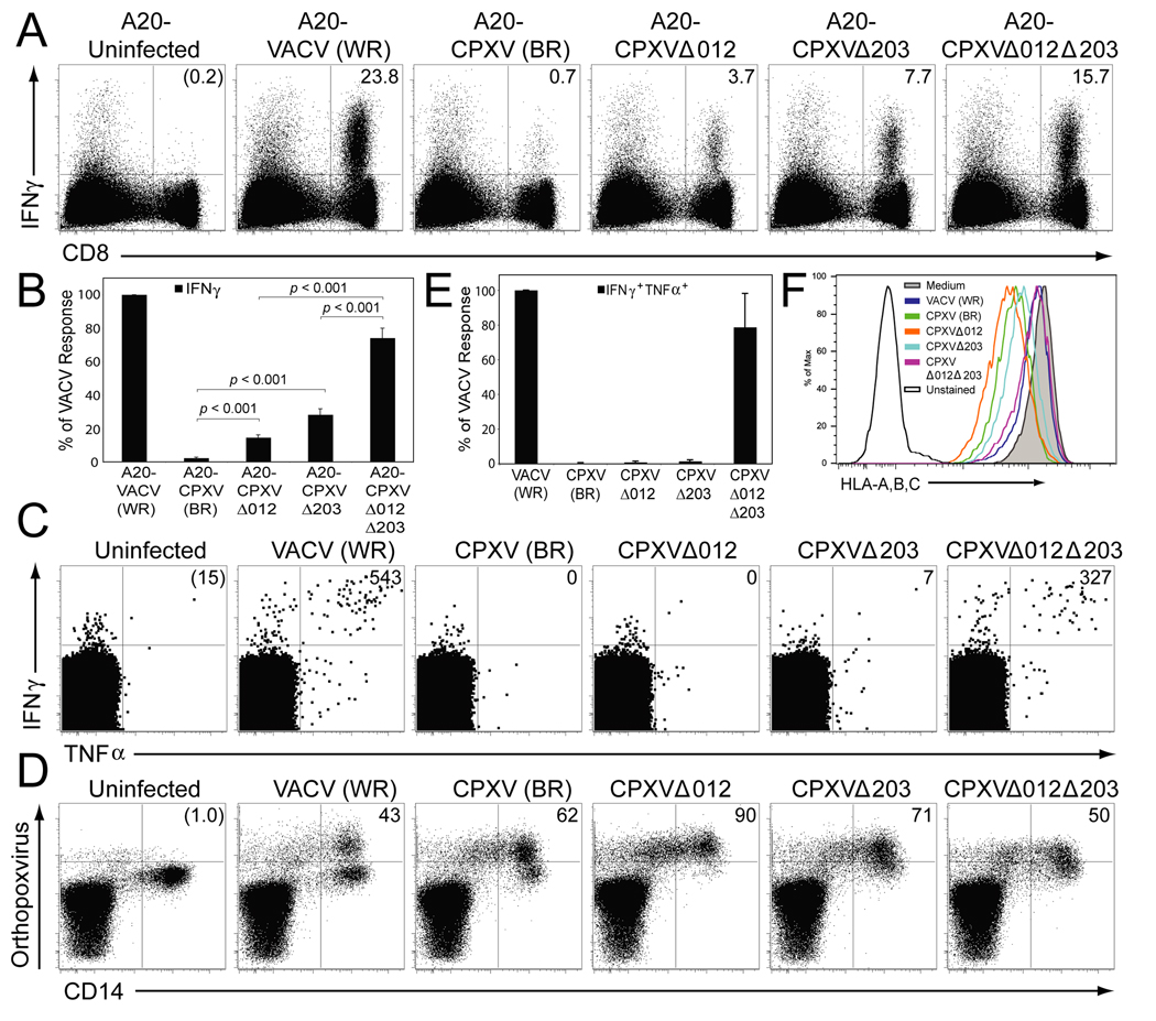
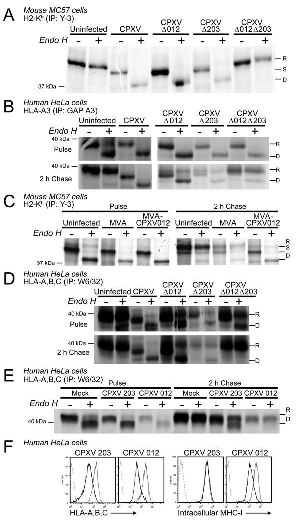
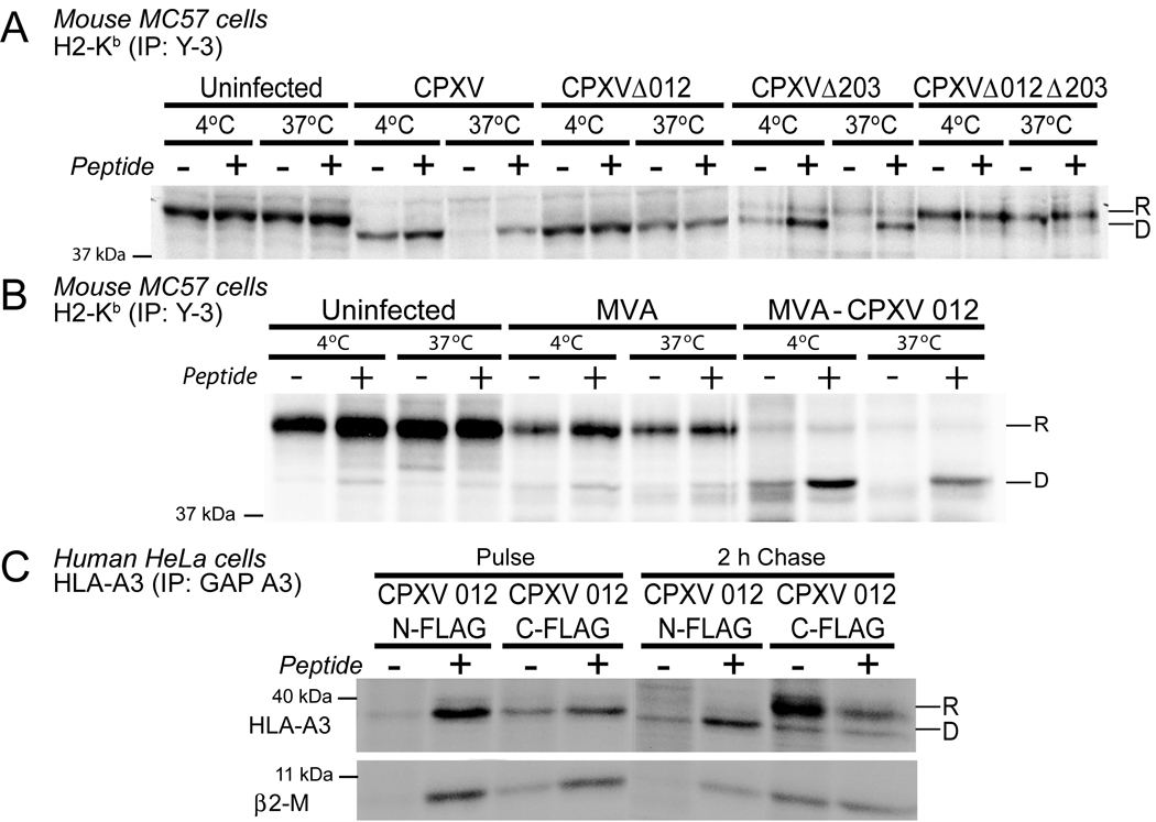
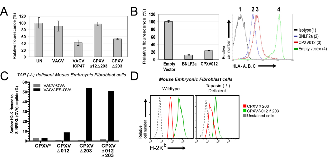
Cowpox virus encodes an extensive array of putative immunomodulatory proteins, likely contributing to its wide host range, which includes zoonotic infections in humans. Unlike Vaccinia virus, cowpox virus prevents stimulation of CD8(+) T cells, a block that correlated with retention of MHC class I in the endoplasmic reticulum by the cowpox virus protein CPXV203. However, deletion of CPXV203 did not restore MHC class I transport or T cell stimulation. Here, we demonstrate the contribution of an additional viral protein, CPXV12, which interferes with MHC class I/peptide complex formation by inhibiting peptide translocation by the transporter associated with antigen processing (TAP). Importantly, human and mouse MHC class I transport and T cell stimulation was restored upon deletion of both CPXV12 and CPXV203, suggesting that these unrelated proteins independently mediate T cell evasion in multiple hosts. CPXV12 is a truncated version of a putative NK cell ligand, indicating that poxviral gene fragments can encode new, unexpected functions.


Conflict of interest statement

The authors declare that they have no conflict of interest.

Figures

Figure 1
Figure 1. CPXV012 downregulates MHC-I surface expression
(A) Flow cytometry of MHC-I surface levels in the presence of CPXV012. HeLa cells were transiently transfected with either pCPXV012 (red) or empty vector (green). At 48 hpt, cells were stained with anti-HLA-A, B, C or anti-CD44 and analyzed by flow-cytometry. Solid grey line represents isotype control. (B) Amino acid alignment of CPXV012 and its orthologs, CPXVD10L (GRI) and CPXVD10L (GER 91). Identical residues are highlighted in yellow. Underlined residues represent the C-type lectin like domain. (C) Flow cytometry of MHC-I surface levels in the presence of CPXV012 and CPXVD10L. pCPXV012-N-FLAG, pCPXV012-C-FLAG, pCPXV012CO-N-FLAG, pCPXVD10L (GER)-N-FLAG or pCPXVD10L (GRI)-N-FLAG were transiently transfected into HeLa cells. The cells were incubated for 48 h and stained with either HLA-specific antibodies (unpermeabilized cells) or anti-FLAG antibodies (permeabilized cells).
Figure 2
Figure 2. CPXV012 is a short lived, ER resident type II transmembrane protein
(A) Model of CPXV012 topology in the ER membrane. (B) In vitro translation of CPXV012 mRNA in the presence of microsomes. In vitro transcribed mRNAs encoding CPXV012 and control proteins, CFP and prolactin were added to a rabbit reticulocyte translation system in the presence of canine microsomal membranes. Left panel: Total translation products or products recovered after microsome pelleting. Right panel: Following translation, pelleted microsomal fractions were treated with proteinase K and separated by SDS-PAGE. Molecular weight markers (kDa) are shown. (C) Subcellular localization of CPXV012. HeLa cells were transiently transfected with pCPXV012-N-FLAG. After 48 h, cells were fixed, permeabilized, and stained with antibodies specific to calreticulin, ERGIC53, giantin, or FLAG. (D) CPXV012 half-life by pulse-chase. pCPXV012-CON-FLAG and empty vector control (Mock) were transiently transfected into HeLa cells. At 24 hpt, cells were pulse-labeled for 1 h and the label was chased for the indicated time. Lysates were immunoprecipitated with anti-FLAG, separated by SDS-PAGE, and visualized by autoradiography (left panel). Optical density of CPXV012-specific bands was measured and plotted against the chase time (right panel).
Figure 3
Figure 3. CPXV012 contributes to MHC-I downregulation by CPXV
(A) Schematic design of Δ012 and Δ012Δ203 mutant viruses. (B) PCR- analysis of Δ012 and Δ012Δ203 mutants. Purified viral genomic DNA of WT CPXV, Δ012 and Δ012Δ203 was PCR amplified with primers specific for CPXV012, CPXV203, or CPXV214. PCR products were separated by gel electrophoresis. (C) Flow cytometry of MHC-I surface expression. THF (left) and MC57 (right) cells were infected with WT CPXV, Δ012, Δ203, and Δ012Δ203. At 24hpi, cells were stained with either HLA- or H2-Kb-specific antibodies.
Figure 4
Figure 4. CD8+ T cell activation by CPXV is restored upon deletion of both CPXV012 and CPXV203
(A) The antiviral CD8+ T cell response in BALB/c mice measured by ICCS. Splenocytes (8 dpi) were incubated with uninfected A20 cells, or infected with the indicated viruses. The percentage of IFNγ producing CD8+ T cells (upper right quadrant) was determined after background subtraction (uninfected control). (B) The average CD8+ T cell response of 6 mice was determined by ICCS and normalized to 100% based on the response to VACV. The data are representative of 2 experiments with 3 mice per group. Statistical differences were calculated using the 2-tailed paired Student t-test. (C) Human virus-specific CD8+ T cell responses in the presence of indicated viruses. PBMC of VACV-immune subjects were gated on CD8+CD4 T cells and the number of IFNγ +TNFα+CD8+ T cells per 106 CD8+ T cells (upper right quadrant) was determined after background subtraction (uninfected control). The CD8+ T cell responses from a representative subject at 2 months after VACV infection is shown. D) The number of virus-infected CD14+ monocytes in a representative PBMC sample at 16 hpi was determined by staining with polyclonal antibodies against OPXV antigens. The numbers in the upper right quadrants represent the percentage of virus-infected CD14+ cells after background subtraction (uninfected). (E) The average antiviral CD8+ T cell response from 4 subjects determined by ICCS at 2 months post-VACV infection and normalized to 100% based on the response to VACV. (F) HLA-A, B, C-expression on virus-infected primary CD14+ monocytes at 16 h p.i.. Monocytes were identified based on forward and side scatter characteristics and CD14 expression.
Figure 5
Figure 5. CPXV012 impaires intracellular transport and steady state levels of MHC-I
(A) H2-Kb expression in the presence of CPXV012. MC57 cells were infected with indicated viruses, pulse labeled for 20 min at 6hpi followed by 2 h chase. Immunoprecipitated samples were EndoH-treated as indicated, separated by SDS-PAGE, and visualized by autoradiography. “R”, “S”, and “D” refer to EndoH-resistant, sensitive, and digested HC. The reduced amount of MHC-I in EndoH-treated samples is due to reduced protein recovery after EndoH treatment. B) Intracellular transport of HLA-A3 transfected into HeLa cells and infected with WT or recombinant CPXV. At 6 hpi, cells were pulse labeled for 20 min followed by 2h chase. HLA-A3 was immunoprecipitated and EndoH-treated as indicated. C) H2-Kb immunoprecipitation from MC57 cells infected with MVA or MVA expressing CPXV012. Cells were labeled and immunoprecipitated as in A. D) Immunoprecipation of endogenous HLA-A,B,C in HeLa cells infected with indicated viruses. Cells were pulse labeled for 20 min at 6h p.i. and chased for 2h. HLA-A,B,C was immunoprecipitated and then treated with EndoH as in A.. E) HeLa cells transiently transfected with CPXV012, CPXV203 or vector control (Mock) were pulse-labeled for 20 min at 24 hpt followed by 2h chase and immunoprecipitation with anti-HLA-A,B,C. F) Intracellular and cell surface levels of MHC-I in the presence of transiently expressed CPXV012 measured by flow cytometry at 48 h after HeLa cells were transfected with pCPXV203 and pCPXV012. Permeabilized cells (intracellular staining) or unfixed cells were stained with anti-HLA-A, B, C. Vector-transfected and isotype control samples are represented by grey and dashed grey lines, respectively.
Figure 6
Figure 6. CPXV012 inhibits peptide loading of MHC-I
(A) and (B) H2-Kb thermostability in the presence of virus expressed CPXV012. MC57 cells were infected with indicated viruses. At 6 hpi, cells were pulse-labeled for 20 min followed by 2 h chase. Lysates were incubated at indicated temperatures in the presence or absence of the peptide SIINFEKEL. H2-Kb was immunoprecipitated with mab Y3 and EndoH-treated. (C). Thermostability of HLA-A3 transfected in HeLa together with CPXV012-N-FLAG or CPXV012-C-FLAG. Cells were labeled as in A and incubated at indicated temperature in the presence or absence of peptide ILRGSVAHK. “R” and “D” refer to EndoH-resistant and digested MHC-I.
Figure 7
Figure 7. CPXV012 inhibits TAP-dependent peptide transport
(A) Peptide transport in the presence of CPXV012. HeLa cells were infected with indicated viruses and at 6 hpi permeabilized with SLO followed by incubation with peptide FITC~CVNKTERAY in the presence or absence of ATP in transport buffer. Recovered fluorescence was background (−ATP) adjusted and is shown as percent relative to uninfected cells. (B) Peptide transport in CPXV012-expressing cells. Peptide transport was measured in MJS cells stably transduced with GFP-expressing control lentiviruses or lentiviruses expressing either BNLF2a or CPXV012 (left panel). Transduction efficiency was verified by monitoring MHC-I surface levels by flow cytometry (right panel). CPXV012, BNLF2a, GFP control, and isotype control are represented by red, blue, green, and black lines, respectively. (C) CPXV012 does not inhibit TAP-independent peptide loading. TAP−/− MEFs were infected with VACV-OVA or VACV-ES-OVA together with indicated CPXV recombinants (MOI=10 each) and simultaneously treated with 25 U/ml IFN-γ. At 24 hpi, SIINFEKL/H2-Kb complexes were measured with specific Ab 25.D1.16 by flow cytometry. The percentage of GFP+ cells that were also 25.D1.16 positive is shown except for WT-CPXV showing total 25.D1.16-positive cells (*). (D) MHC-I downregulation by CPXV012 does not require Tapasin. WT and TPN−/− MEFs were infected with indicated viruses (MOI=5) in the presence of IFN-γ and surface H2-Kb was monitored by flow cytometry at 24 hpi. Kb-expression is shown for GFP+ cells.

Comment in

Similar articles

Cited by

References

    1. Ahn K, Meyer TH, Uebel S, Sempe P, Djaballah H, Yang Y, Peterson PA, Früh K, Tampe R. Molecular mechanism and species specificity of TAP inhibition by herpes simplex virus ICP47. Embo J. 1996;15:3247–3255. - PMC - PubMed
    1. Bacik I, Cox JH, Anderson R, Yewdell JW, Bennink JR. TAP (transporter associated with antigen processing)-independent presentation of endogenously synthesized peptides is enhanced by endoplasmic reticulum insertion sequences located at the amino- but not carboxyl-terminus of the peptide. J Immunol. 1994;152:381–387. - PubMed
    1. Bakker AH, Hoppes R, Linnemann C, Toebes M, Rodenko B, Berkers CR, Hadrup SR, van Esch WJ, Heemskerk MH, Ovaa H, et al. Conditional MHC class I ligands and peptide exchange technology for the human MHC gene products HLA-A1, -A3, - A11, and -B7. Proc Natl Acad Sci U S A. 2008;105:3825–3830. - PMC - PubMed
    1. Banks TA, Jenkins FJ, Kanangat S, Nair S, Dasgupta S, Foster CM, Rouse BT. Vaccination with the immediate-early protein ICP47 of herpes simplex virus-type 1 (HSV-1) induces virus-specific lymphoproliferation, but fails to protect against lethal challenge. Virology. 1994;200:236–245. - PubMed
    1. Barel MT, Pizzato N, Le Bouteiller P, Wiertz EJ, Lenfant F. Subtle sequence variation among MHC class I locus products greatly influences sensitivity to HCMV US2- and US11-mediated degradation. International immunology. 2006;18:173–182. - PubMed

Publication types

MeSH terms

Substances