. 2010 Jan 8;285(2):914-22.
doi: 10.1074/jbc.M109.010892. Epub 2009 Nov 16.

Self-association of calcium-binding protein S100A4 and metastasis

Affiliations

Self-association of calcium-binding protein S100A4 and metastasis

Thamir M Ismail et al. J Biol Chem. .

Abstract

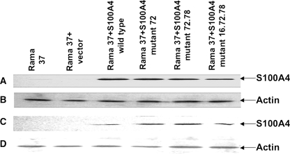
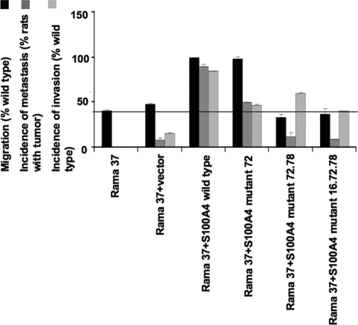
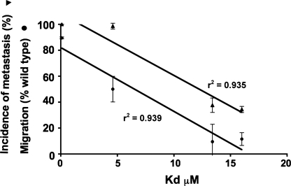
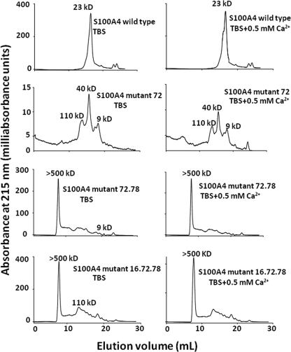
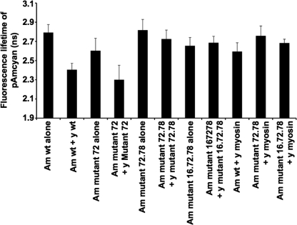
Elevated levels of the calcium-binding protein S100A4 promote metastasis and in carcinoma cells are associated with reduced survival of cancer patients. S100A4 interacts with target proteins that affect a number of activities associated with metastatic cells. However, it is not known how many of these interactions are required for S100A4-promoted metastasis, thus hampering the design of specific inhibitors of S100A4-induced metastasis. Intracellular S100A4 exists as a homodimer through previously identified, well conserved, predominantly hydrophobic key contacts between the subunits. Here it is shown that mutating just one key residue, phenylalanine 72, to alanine is sufficient to reduce the metastasis-promoting activity of S100A4 to 50% that of the wild type protein, and just 2 or 3 specific mutations reduces the metastasis-promoting activity of S100A4 to less than 20% that of the wild type protein. These mutations inhibit the self-association of S100A4 in vivo and reduce markedly the affinity of S100A4 for at least two of its protein targets, a recombinant fragment of non-muscle myosin heavy chain isoform A, and p53. Inhibition of the self-association of S100 proteins might be a novel means of inhibiting their metastasis-promoting activities.


Figures

FIGURE 1.
FIGURE 1.
The level of wild type and mutant S100A4 proteins in transfected Rama 37 cell clones and cell pools. Cells were transfected with expression vectors containing S100A4 wild type or mutant 72, 72.78, or 16.72.78 cDNAs as described under “Experimental Procedures.” Extracts of cloned cell lines (A and B) or pooled transfectants (C and D) were separated by SDS-PAGE and blotted onto polyvinylidene difluoride membranes. Membranes were incubated with either a polyclonal S100A4 antibody (A and C) or anti-actin serum (B and D) as a loading control. Bound antibodies were detected by chemiluminescence with horseradish peroxidase-conjugated secondary antibody as described previously (28, 30, 31). The intensities of the bands of S100A4 protein corrected for actin levels in cloned cell lines, Rama 37, Rama 37 + vector, Rama 37 + S100A4 mutant 72, Rama 37 + S100A4 mutant 72.78, and Rama 37 + S100A4 mutant 16.72.78 were 16, 10, 95, 90, and 88% that of wild type and, for cell pools, 10, 5, 99, 110, and 91% that of wild type.
FIGURE 2.
FIGURE 2.
The effects of interface mutations of S100A4 on metastasis and invasion in vivo and S100A4-induced migration of Rama 37 cells in vitro. Cell pools or clones of Rama 37 cells expressing wild type S100A4 or mutants 72, 72.78, or 16.72.78 were subjected to migration assays in quadruplicate Transwell chambers as described under “Experimental Procedures.” Parental Rama 37 cell line and cells transfected with empty vector were used as negative controls. For statistical analysis, at least four independent assays were conducted on clones and four on pools of cells. The results were converted to a percentage of wild type migration. The horizontal line denotes the basal level of migration of the Rama 37 cells in the Transwell assay. Incidences of metastasis and invasion are expressed as the percentages of animals with primary tumors using data from Table 1.
FIGURE 3.
FIGURE 3.
Relationship between motility, metastasis, and the interaction of rNMMIIHCA with wild type and mutant S100A4 proteins. The equilibrium dissociation constants for the interaction of mutant S100A4 proteins and immobilized rNMMIIHCA were determined using an optical biosensor and plotted against the motility and metastasis-promoting properties of Rama 37 cells expressing wild type or mutant S100A4 proteins.
FIGURE 4.
FIGURE 4.
Elution profile of S100A4 wild type and mutant proteins on gel filtration chromatography. Samples were applied to a Superdex 200 10/300 GL column equilibrated with TBS in the absence of (left-hand column) or in the presence of 0.5 mm Ca2+ (right-hand column). Proteins were separated at a flow rate of 0.5 ml/min, and elution was monitored at 215 nm. The retention times were related to those of standard proteins (“Experimental Procedures”) to determine the molecular masses of the S100A4 wild type and mutant proteins.
FIGURE 5.
FIGURE 5.
FLIM-FRET analysis. Fluorescence lifetime imaging of cells expressing AmCyan-S100A4, YFP-S100A4, and myosin-YFP proteins are shown. The interaction at the level of one living cell was measured using laser scanning microscopy as described under “Experimental Procedures.” Cells expressing AmCyan-wild type S100A4 and YFP-wild type S100A4 (7 cells were measured) and cells expressing AmCyan-S100A4 mutant 72 protein and YFP-S100A4 mutant 72 protein (4 cells were measured) exhibited significantly reduced fluorescence lifetime compared with control cells expressing AmCyan-wild type S100A4 alone (9 cells were measured) or AmCyan mutant 72 protein alone (5 cells were measured). In contrast, cells expressing AmCyan-S100A4 mutant 72.78 and YFP-S100A4 mutant 72.78 (11 cells were measured) or AmCyan-S100A4 mutant 16.72.78 and YFP-S100A4 mutant 16.72.78 (5 cells were measured) did not show reduced fluorescence lifetimes relative to controls expressing AmCyan-S100A4 mutant 72.78 (11 cells were measured) or AmCyan-S100A4 mutant 16.72.78 alone (9 cells were measured). Am, AmCyan fluorescent protein; y, yellow fluorescent protein; wt, wild type.

Similar articles

Cited by

References

    1. Andersen K., Nesland J. M., Holm R., Flørenes V. A., Fodstad Ø., Maelandsmo G. M. (2004) Mod. Pathol. 17, 990–997 - PubMed
    1. Kimura K., Endo Y., Yonemura Y., Heizmann C. W., Schafer B. W., Watanabe Y., Sasaki T. (2000) Int. J. Oncol. 16, 1125–1131 - PubMed
    1. Matsubara D., Niki T., Ishikawa S., Goto A., Ohara E., Yokomizo T., Heizmann C. W., Aburatani H., Moriyama S., Moriyama H., Nishimura Y., Funata N., Fukayama M. (2005) Cancer Sci. 96, 844–857 - PMC - PubMed
    1. Rudland P. S., Platt-Higgins A., Renshaw C., West C. R., Winstanley J. H., Robertson L., Barraclough R. (2000) Cancer Res. 60, 1595–1603 - PubMed
    1. Lee W. Y., Su W. C., Lin P. W., Guo H. R., Chang T. W., Chen H. H. (2004) Oncology 66, 429–438 - PubMed

Publication types

MeSH terms