. 2010 Jan 15;285(3):1879-87.
doi: 10.1074/jbc.M109.055392. Epub 2009 Nov 16.

The protein kinase Cdelta catalytic fragment is critical for maintenance of the G2/M DNA damage checkpoint

Affiliations

The protein kinase Cdelta catalytic fragment is critical for maintenance of the G2/M DNA damage checkpoint

Edward L LaGory et al. J Biol Chem. .

Abstract

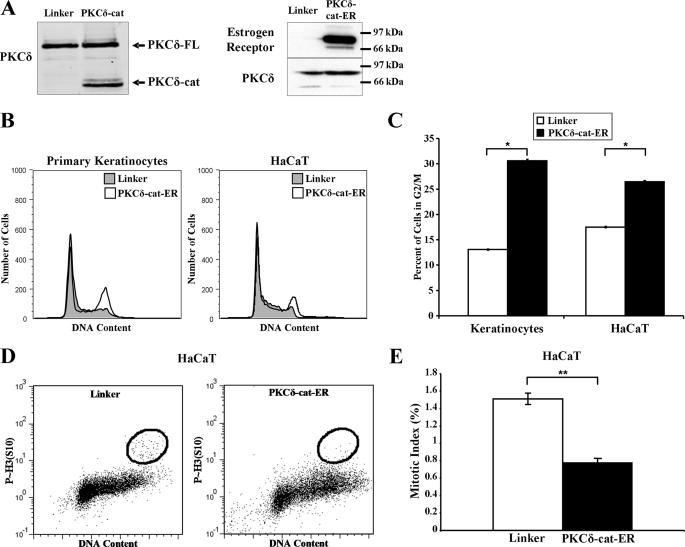
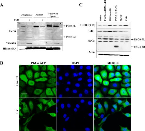
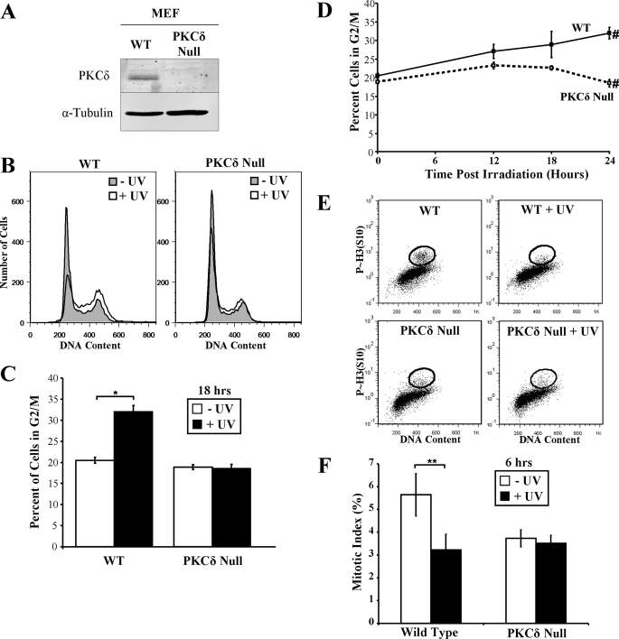
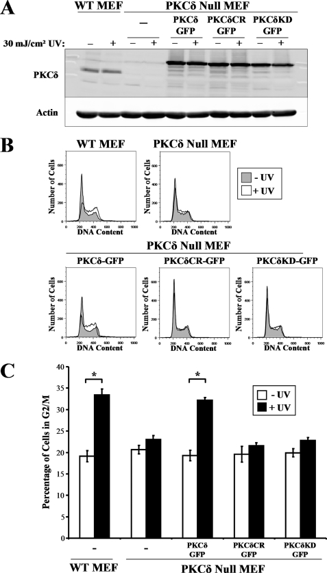
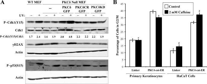
Protein kinase Cdelta (PKCdelta) is an essential component of the intrinsic apoptotic program. Following DNA damage, such as exposure to UV radiation, PKCdelta is cleaved in a caspase-dependent manner, generating a constitutively active catalytic fragment (PKCdelta-cat), which is necessary and sufficient for keratinocyte apoptosis. We found that in addition to inducing apoptosis, expression of PKCdelta-cat caused a pronounced G(2)/M cell cycle arrest in both primary human keratinocytes and immortalized HaCaT cells. Consistent with a G(2)/M arrest, PKCdelta-cat induced phosphorylation of Cdk1 (Tyr(15)), a critical event in the G(2)/M checkpoint. Treatment with the ATM/ATR inhibitor caffeine was unable to prevent PKCdelta-cat-induced G(2)/M arrest, suggesting that PKCdelta-cat is functioning downstream of ATM/ATR in the G(2)/M checkpoint. To better understand the role of PKCdelta and PKCdelta-cat in the cell cycle response to DNA damage, we exposed wild-type and PKCdelta null mouse embryonic fibroblasts (MEFs) to UV radiation. Wild-type MEFs underwent a pronounced G(2)/M arrest, Cdk1 phosphorylation, and induction of apoptosis following UV exposure, whereas PKCdelta null MEFs were resistant to these effects. Expression of PKCdelta-green fluorescent protein, but not caspase-resistant or kinase-inactive PKCdelta, was able to restore G(2)/M checkpoint integrity in PKCdelta null MEFs. The function of PKCdelta in the DNA damage-induced G(2)/M cell cycle checkpoint may be a critical component of its tumor suppressor function.


Figures

FIGURE 1.
FIGURE 1.
PKCδ-cat expression induces G2/M cell cycle arrest. A, Western blots displaying relative levels of full-length PKCδ protein (PKCδ-FL) and PKCδ-cat are shown in control Linker-, PKCδ-cat-FLAG-, and PKCδ-cat-ER-transduced KCs. PKCδ-cat-ER-transduced cells display a strong ER-positive band at the same position as the PKCδ protein, indicating successful expression of the ER fusion protein. B, representative DNA content histograms of primary KCs and HaCaT cells transduced with either Linker or PKCδ-cat-ER retrovirus are shown. Propidium iodide staining was performed 2–3 days after infection. Similar results were obtained in at least three independent experiments. C, quantitation of the percentage of cells containing G2/M DNA content in KCs and HaCaT cells 2–3 days following infection with either Linker control or PKCδ-cat-ER retrovirus. Graphs represent experiments done in triplicate. *, Student's t test value of p < 0.005. D, HaCaT cells transduced with either Linker or PKCδ-cat-ER retrovirus were harvested and stained for phosphorylated histone H3 (Ser10) and propidium iodide after 3 days and analyzed by flow cytometry. E, quantitation of mitotic indices from HaCaT cells treated as described for D. **, Student's t test value of p < 0.001. Error bars denote the S.D.
FIGURE 2.
FIGURE 2.
PKCδ-cat expression induces G2/M checkpoint activation in KCs. A, Western blot showing levels of PKCδ protein in cytoplasmic and nuclear extracts of untreated or 30 mJ/cm2 UV radiation-exposed KCs after 18 h. Full-length PKCδ (PKCδ-FL) and PKCδ-cat are indicated by arrows. Vinculin and histone H3 are presented as cytoplasmic and nuclear markers, respectively. B, confocal microscope images were taken showing cytoplasmic and nuclear staining of PKCδ-GFP fusion protein both before and 18 h after exposure to 30 mJ/cm2 UV radiation. 4′,6-Diamidino-2-phenylindole (DAPI) staining is displayed to demonstrate nuclear localization. Images were taken using a ×40 objective and 1-μm optical slice. Scale bar denotes 25 μm. C, Western blot showing levels of p-Cdk1 (Tyr15), total Cdk1, and PKCδ after retroviral transduction of PKCδ-cat-ER, PKCδ-cat, or the kinase-dead PKCδ(K378A)-cat-ER. The PKCδ-cat-ER fusion proteins run to the same position in the gel as the full-length PKCδ. KC lysate harvested 18 h after exposure to 30 mJ/cm2 UV radiation is shown as a positive control for p-Cdk1 (Tyr15) induction. Actin protein levels are displayed as a loading control.
FIGURE 3.
FIGURE 3.
PKCδ null MEFs fail to arrest in G2/M phase following UV irradiation. A, Western blot displaying PKCδ protein levels in wild-type (WT) and PKCδ null MEF whole cell lysates. α-Tubulin levels are shown as a loading control. B, representative DNA content histograms of wild-type and PKCδ null MEFs before and 18 h after exposure to 30 mJ/cm2 UV radiation. C, the percentage of wild-type and PKCδ null MEFs in G2/M phase of the cell cycle before and after exposure to 30 mJ/cm2 UV radiation is displayed. Error bars denote the S.D. from experiments performed in triplicate. *, Student's t test value of p < 0.005. D, the percentage of wild-type and PKCδ null MEFs with G2/M DNA content at various times following exposure to 30 mJ/cm2 UV radiation is displayed. Error bars denote the S.D. from experiments performed in triplicate. #, Student's t test value of p < 0.005. E, wild-type and PKCδ null MEFs were exposed to 10 mJ/cm2 UV radiation, treated with 100 ng/ml nocodazole, and stained for phosphorylated histone H3 (Ser10) and propidium iodide 6 h after UV exposure. Flow cytometry analysis is shown, with the phosphorylated histone H3 (Ser10)-positive G2/M DNA content cells circled as the mitotic cells. F, quantitation of mitotic indices from HaCaT cells treated as described for D. **, Student's t test value of p < 0.05.
FIGURE 4.
FIGURE 4.
G2/M checkpoint induction requires PKCδ kinase activity. A, wild-type (WT) and PKCδ null MEFs were untransduced or transduced with wild-type PKCδ-GFP, cleavage-resistant (CR) PKCδ(D327A)-GFP, or kinase-dead (KD) PKCδ(K376R)-GFP and exposed to 30 mJ/cm2 UV radiation. Protein lysates were analyzed for expression of PKCδ and actin. B, representative DNA content histograms of wild-type and PKCδ null MEFs before and 18 h after exposure to 30 mJ/cm2 UV radiation. C, the percentage of wild-type and PKCδ null MEFs in G2/M after transduction with the indicated PKCδ-GFP fusion protein before and after exposure to 30 mJ/cm2 UV radiation is displayed. Error bars denote the S.D. from experiments performed in triplicate. *, Student's t test value of p < 0.005.
FIGURE 5.
FIGURE 5.
UV radiation-induced G2/M DNA damage checkpoint activation requires PKCδ function. A, wild-type (WT) and PKCδ null MEFs expressing PKCδ-GFP, cleavage-resistant (CR) PKCδ(D327A)-GFP, or kinase-dead (KD) PKCδ(K376R)-GFP were exposed to 10 mJ/cm2 UV radiation. Protein lysates were collected 18 h after irradiation and analyzed for levels of p-Cdk1 (Tyr15), total Cdk1, γH2A.X, and actin. p-Cdk1 (Tyr15)/Cdk1 densitometry ratios are displayed and were similar in multiple experiments. B, propidium iodide cell cycle analysis of LZRS-Linker- or LZRS-PKCδ-cat-ER-transduced primary KCs and HaCaT cells incubated with 2 mm caffeine for 48 h. The percentage of cells in the G2/M phase of the cell cycle is displayed from experiments performed in triplicate. *, Student's t test value of p < 10−5; #, Student's t test value of p < 10−4.

References

    1. Bartek J., Lukas J. (2007) Curr. Opin. Cell Biol. 19, 238–245 - PubMed
    1. Musacchio A., Salmon E. D. (2007) Nat. Rev. Mol. Cell Biol. 8, 379–393 - PubMed
    1. Hollstein M., Sidransky D., Vogelstein B., Harris C. C. (1991) Science 253, 49–53 - PubMed
    1. Liu Q., Guntuku S., Cui X. S., Matsuoka S., Cortez D., Tamai K., Luo G., Carattini-Rivera S., DeMayo F., Bradley A., Donehower L. A., Elledge S. J. (2000) Genes Dev. 14, 1448–1459 - PMC - PubMed
    1. Banin S., Moyal L., Shieh S., Taya Y., Anderson C. W., Chessa L., Smorodinsky N. I., Prives C., Reiss Y., Shiloh Y., Ziv Y. (1998) Science 281, 1674–1677 - PubMed

Publication types

Substances