

A sensitive fluorescence-based assay for monitoring GM2 ganglioside hydrolysis in live patient cells and their lysates
- PMID: 19917668
- PMCID: PMC2896968
- DOI: 10.1093/glycob/cwp183
A sensitive fluorescence-based assay for monitoring GM2 ganglioside hydrolysis in live patient cells and their lysates
Abstract
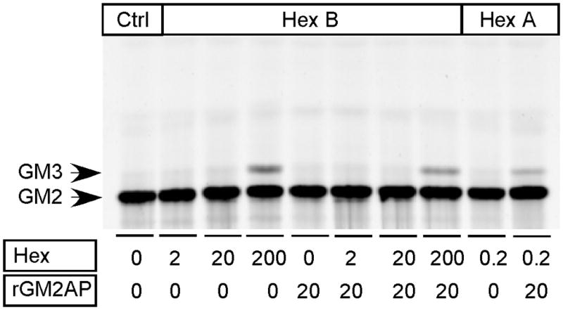
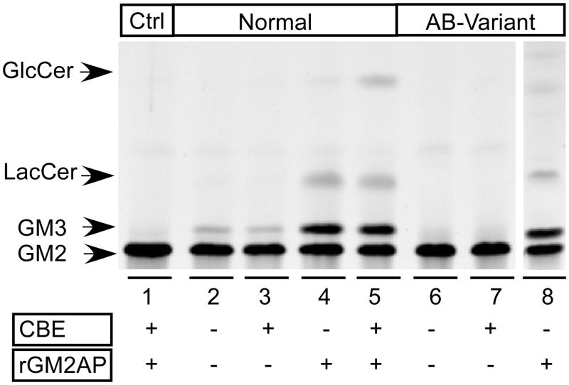
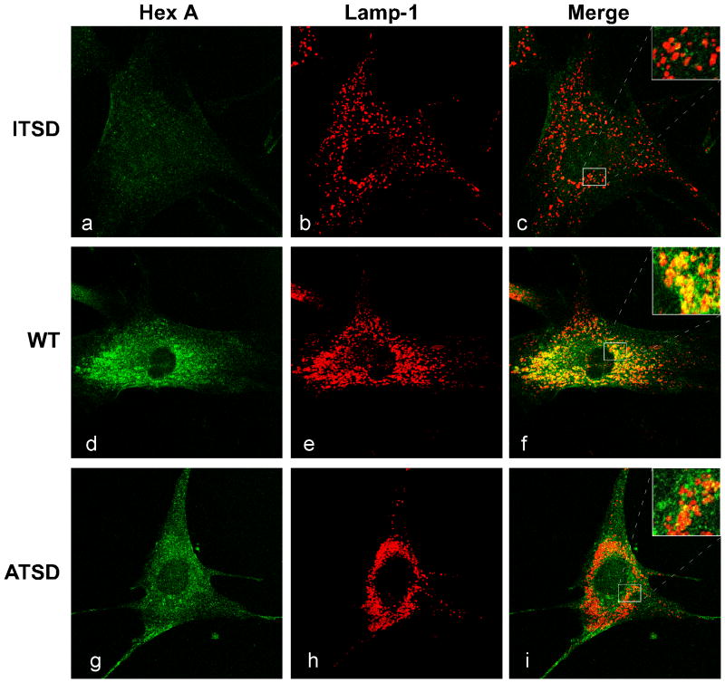
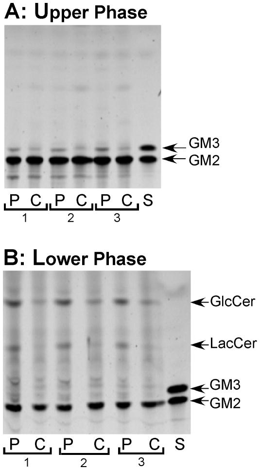
Enzyme enhancement therapy, utilizing small molecules as pharmacological chaperones, is an attractive approach for the treatment of lysosomal storage diseases that are associated with protein misfolding. However, pharmacological chaperones are also inhibitors of their target enzyme. Thus, a major concern with this approach is that, despite enhancing protein folding within, and intracellular transport of the functional mutant enzyme out of the endoplasmic reticulum, the chaperone will continue to inhibit the enzyme in the lysosome, preventing substrate clearance. Here we demonstrate that the in vitro hydrolysis of a fluorescent derivative of lyso-GM2 ganglioside, like natural GM2 ganglioside, is specifically carried out by the beta-hexosaminidase A isozyme, requires the GM2 activator protein as a co-factor, increases when the derivative is incorporated into anionic liposomes and follows similar Michaelis-Menten kinetics. This substrate can also be used to differentiate between lysates from normal and GM2 activator-deficient cells. When added to the growth medium of cells, the substrate is internalized and primarily incorporated into lysosomes. Utilizing adult Tay-Sachs fibroblasts that have been pre-treated with the pharmacological chaperone Pyrimethamine and subsequently loaded with this substrate, we demonstrate an increase in both the levels of mutant beta-hexosaminidase A and substrate-hydrolysis as compared to mock-treated cells.
Figures


Similar articles
-
In cellulo examination of a beta-alpha hybrid construct of beta-hexosaminidase A subunits, reported to interact with the GM2 activator protein and hydrolyze GM2 ganglioside.PLoS One. 2013;8(3):e57908. doi: 10.1371/journal.pone.0057908. Epub 2013 Mar 4. PLoS One. 2013. PMID: 23483939 Free PMC article.
-
Membrane lipids regulate ganglioside GM2 catabolism and GM2 activator protein activity.J Lipid Res. 2015 Sep;56(9):1747-61. doi: 10.1194/jlr.M061036. Epub 2015 Jul 14. J Lipid Res. 2015. PMID: 26175473 Free PMC article.
-
Lyso-GM2 ganglioside: a possible biomarker of Tay-Sachs disease and Sandhoff disease.PLoS One. 2011;6(12):e29074. doi: 10.1371/journal.pone.0029074. Epub 2011 Dec 20. PLoS One. 2011. PMID: 22205997 Free PMC article.
-
The GM2 activator protein, its roles as a co-factor in GM2 hydrolysis and as a general glycolipid transport protein.Biochim Biophys Acta. 1998 Jul 31;1393(1):1-18. doi: 10.1016/s0005-2760(98)00057-5. Biochim Biophys Acta. 1998. PMID: 9714704 Review.
-
Biochemical consequences of mutations causing the GM2 gangliosidoses.Biochim Biophys Acta. 1999 Oct 8;1455(2-3):105-38. doi: 10.1016/s0925-4439(99)00074-5. Biochim Biophys Acta. 1999. PMID: 10571007 Review.
Cited by
-
Chaperone therapy for GM2 gangliosidosis: effects of pyrimethamine on β-hexosaminidase activity in Sandhoff fibroblasts.Mol Neurobiol. 2014 Aug;50(1):159-67. doi: 10.1007/s12035-013-8605-5. Epub 2013 Dec 20. Mol Neurobiol. 2014. PMID: 24356898 Review.
-
Biochemical Correction of GM2 Ganglioside Accumulation in AB-Variant GM2 Gangliosidosis.Int J Mol Sci. 2023 May 24;24(11):9217. doi: 10.3390/ijms24119217. Int J Mol Sci. 2023. PMID: 37298170 Free PMC article.
-
Lysosomal storage diseases--the horizon expands.Nat Rev Neurol. 2013 Oct;9(10):583-98. doi: 10.1038/nrneurol.2013.163. Epub 2013 Aug 13. Nat Rev Neurol. 2013. PMID: 23938739 Review.
-
In cellulo examination of a beta-alpha hybrid construct of beta-hexosaminidase A subunits, reported to interact with the GM2 activator protein and hydrolyze GM2 ganglioside.PLoS One. 2013;8(3):e57908. doi: 10.1371/journal.pone.0057908. Epub 2013 Mar 4. PLoS One. 2013. PMID: 23483939 Free PMC article.
-
Characterization of the biosynthesis, processing and kinetic mechanism of action of the enzyme deficient in mucopolysaccharidosis IIIC.PLoS One. 2011;6(9):e24951. doi: 10.1371/journal.pone.0024951. Epub 2011 Sep 21. PLoS One. 2011. PMID: 21957468 Free PMC article.
References
-
- Callahan JW, Pinsky L, Wolfe LS. GM1 -gangliosidosis (Type II): studies on a fibroblast cell strain. Biochemical medicine. 1970;4:295–316. - PubMed
-
- Folch J, Lees M, Sloane Stanley GH. A simple method for the isolation and purification of total lipides from animal tissues. J Biol Chem. 1957;226:497–509. - PubMed
-
- Gravel RA, Clarke JTR, Kaback MM, Mahuran D, Sandhoff K, Suzuki K. The GM2 gangliosidoses. In: Scriver CR, Beaudet AL, et al., editors. The Metabolic and Molecular Bases of Inherited Disease. McGraw-Hill; New York: 1995. pp. 2839–2879.
-
- Hou Y, Tse R, Mahuran DJ. The Direct Determination of the Substrate Specificity of the α-Active site in Heterodimeric β-Hexosaminidase A. Biochemistry. 1996;35:3963–3969. - PubMed
Publication types
MeSH terms
Substances
Grants and funding
LinkOut - more resources
Full Text Sources
Other Literature Sources
